Copyright
©The Author(s) 2021.
World J Clin Cases. Apr 16, 2021; 9(11): 2671-2678
Published online Apr 16, 2021. doi: 10.12998/wjcc.v9.i11.2671
Published online Apr 16, 2021. doi: 10.12998/wjcc.v9.i11.2671
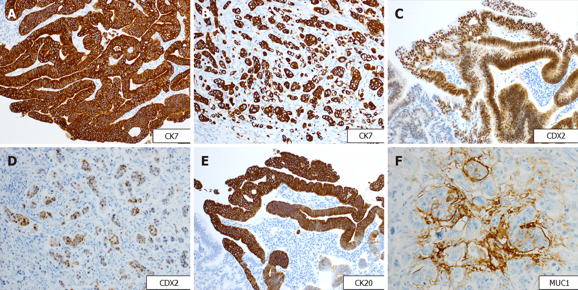
Figure 3 Immunohistochemical findings.
A and B: Immunohistochemical staining for CK7 revealing positivity in both tubular and micropapillary carcinoma (immunostain, magnification 200 ×); C and D: Tumor cells in both tubular and micropapillary portions showing positivity for caudal-related homeobox transcription factor-2 (immunostain, magnification 200 ×); E: Tumor cells on the surface area showing positivity for CK20 (immunostain, magnification 200 ×); F: The micropapillary component showing focally positivity for mucin1 (immunostain, magnification 400 ×). CDX2: Caudal-related homeobox transcription factor-2; MUC1: Mucin1.
- Citation: Noguchi H, Higashi M, Idichi T, Kurahara H, Mataki Y, Tasaki T, Kitazono I, Ohtsuka T, Tanimoto A. Rare histological subtype of invasive micropapillary carcinoma in the ampulla of Vater: A case report. World J Clin Cases 2021; 9(11): 2671-2678
- URL: https://www.wjgnet.com/2307-8960/full/v9/i11/2671.htm
- DOI: https://dx.doi.org/10.12998/wjcc.v9.i11.2671


