Copyright
©The Author(s) 2021.
World J Clin Cases. Apr 16, 2021; 9(11): 2671-2678
Published online Apr 16, 2021. doi: 10.12998/wjcc.v9.i11.2671
Published online Apr 16, 2021. doi: 10.12998/wjcc.v9.i11.2671
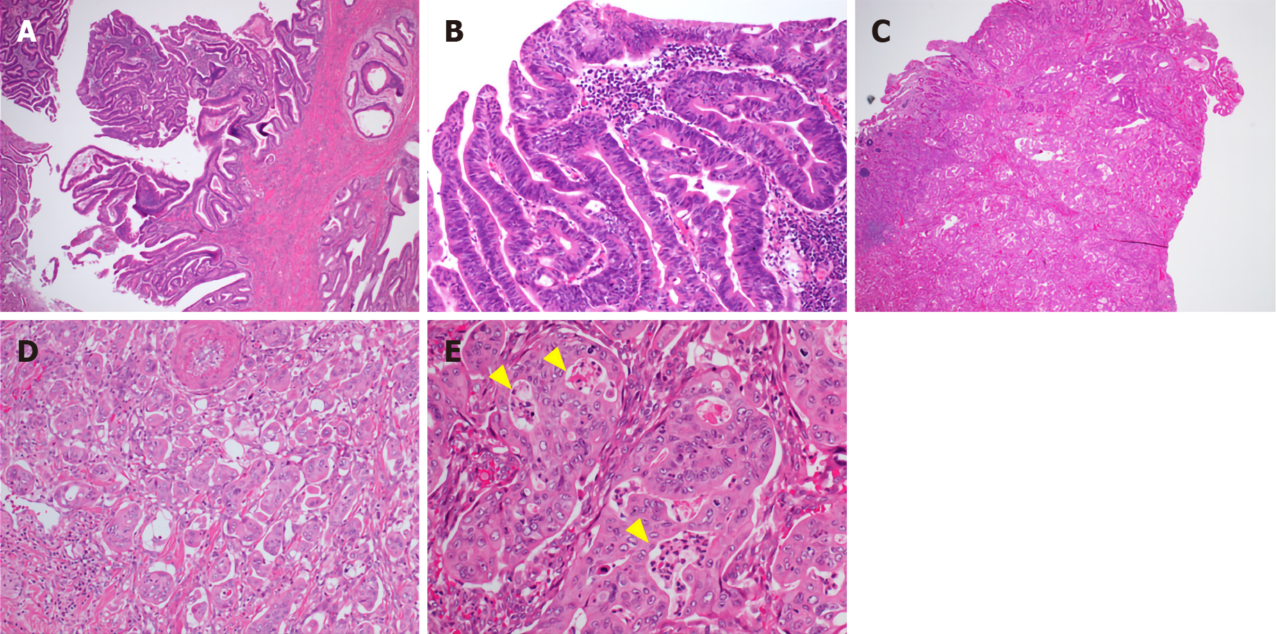
Figure 2 Microscopic findings.
A: The section shows that the surface of the tumor is composed of tubulo-papillary proliferation [hematoxylin and eosin (H&E) stain, magnification 40 ×]; B: Significant architectural and nuclear atypia, including loss of polarity (H&E stain, magnification 200 ×); C: Transition from the tubular structure to the invasive micropapillary pattern (H&E stain, magnification 40 ×); D: The invasion edge of the tumor composed of moderately differentiated adenocarcinoma and micropapillary carcinoma (H&E stain, magnification 200 ×); E: Neutrophilic infiltration forming intraepithelial microabscesses in the tumor nest (H&E stain, magnification 400 ×).
- Citation: Noguchi H, Higashi M, Idichi T, Kurahara H, Mataki Y, Tasaki T, Kitazono I, Ohtsuka T, Tanimoto A. Rare histological subtype of invasive micropapillary carcinoma in the ampulla of Vater: A case report. World J Clin Cases 2021; 9(11): 2671-2678
- URL: https://www.wjgnet.com/2307-8960/full/v9/i11/2671.htm
- DOI: https://dx.doi.org/10.12998/wjcc.v9.i11.2671


