Copyright
©The Author(s) 2021.
World J Clin Cases. Apr 16, 2021; 9(11): 2662-2670
Published online Apr 16, 2021. doi: 10.12998/wjcc.v9.i11.2662
Published online Apr 16, 2021. doi: 10.12998/wjcc.v9.i11.2662
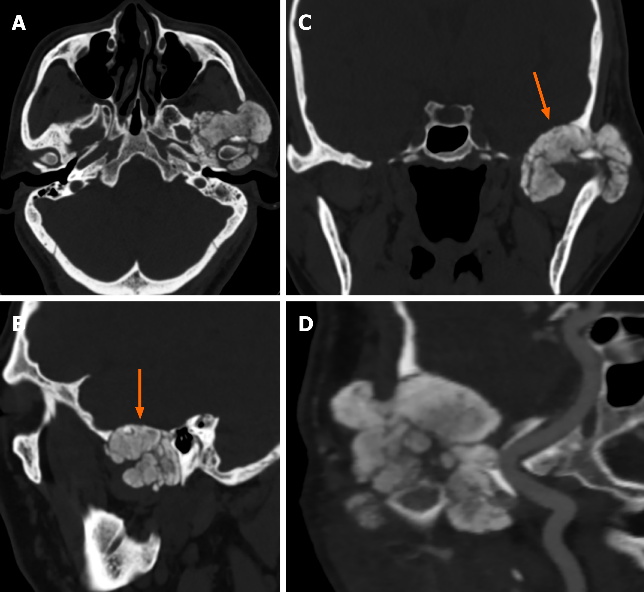
Figure 3 Computed tomography images of Case 2.
A calcified mass in the left temporomandibular joint was shown via computed tomography (A). The mass infiltrated the middle cranial fossa by destroying the skull base (B and C, arrow) and was adjacent to the left internal carotid artery (D).
- Citation: Tang T, Han FG. Calcium pyrophosphate deposition disease of the temporomandibular joint invading the middle cranial fossa: Two case reports. World J Clin Cases 2021; 9(11): 2662-2670
- URL: https://www.wjgnet.com/2307-8960/full/v9/i11/2662.htm
- DOI: https://dx.doi.org/10.12998/wjcc.v9.i11.2662


