Copyright
©The Author(s) 2021.
World J Clin Cases. Apr 16, 2021; 9(11): 2662-2670
Published online Apr 16, 2021. doi: 10.12998/wjcc.v9.i11.2662
Published online Apr 16, 2021. doi: 10.12998/wjcc.v9.i11.2662
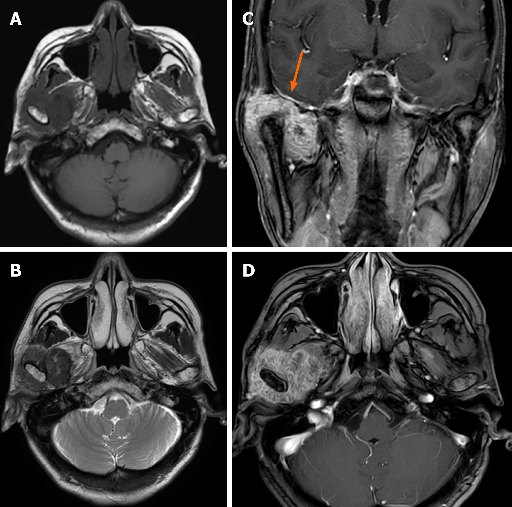
Figure 2 Magnetic resonance imaging images of Case 1.
Axial T1-weighted image (A) and T2-weighted image (B) showed a hypointense mass. The lesion was markedly enhanced after enhancement (C and D) and in contact with the dura mater on the inferior surface (C, arrow).
- Citation: Tang T, Han FG. Calcium pyrophosphate deposition disease of the temporomandibular joint invading the middle cranial fossa: Two case reports. World J Clin Cases 2021; 9(11): 2662-2670
- URL: https://www.wjgnet.com/2307-8960/full/v9/i11/2662.htm
- DOI: https://dx.doi.org/10.12998/wjcc.v9.i11.2662


