Copyright
©The Author(s) 2021.
World J Clin Cases. Apr 6, 2021; 9(10): 2380-2385
Published online Apr 6, 2021. doi: 10.12998/wjcc.v9.i10.2380
Published online Apr 6, 2021. doi: 10.12998/wjcc.v9.i10.2380
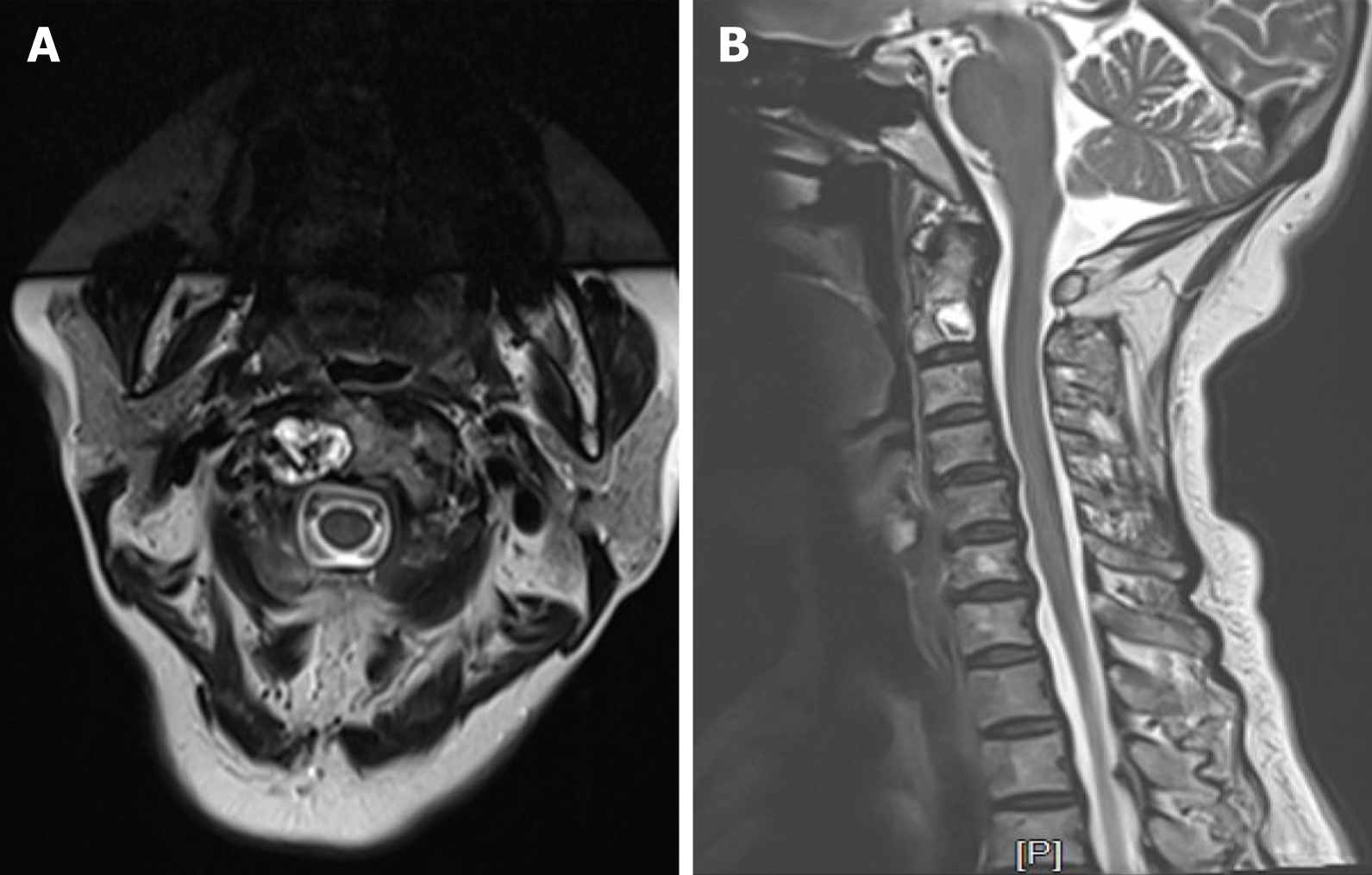
Figure 3 Magnetic resonance imaging.
A: A high signal mass on the right side of C2 with an irregular boundary; B: Cervical disc herniation could be found in C4-6, while no garrulous blade or swollen discs were observed toward the spinal cord in C2.
- Citation: Li RJ, Li XF, Jiang WM. Solitary bone plasmacytoma of the upper cervical spine: A case report . World J Clin Cases 2021; 9(10): 2380-2385
- URL: https://www.wjgnet.com/2307-8960/full/v9/i10/2380.htm
- DOI: https://dx.doi.org/10.12998/wjcc.v9.i10.2380


