Copyright
©The Author(s) 2021.
World J Clin Cases. Apr 6, 2021; 9(10): 2367-2372
Published online Apr 6, 2021. doi: 10.12998/wjcc.v9.i10.2367
Published online Apr 6, 2021. doi: 10.12998/wjcc.v9.i10.2367
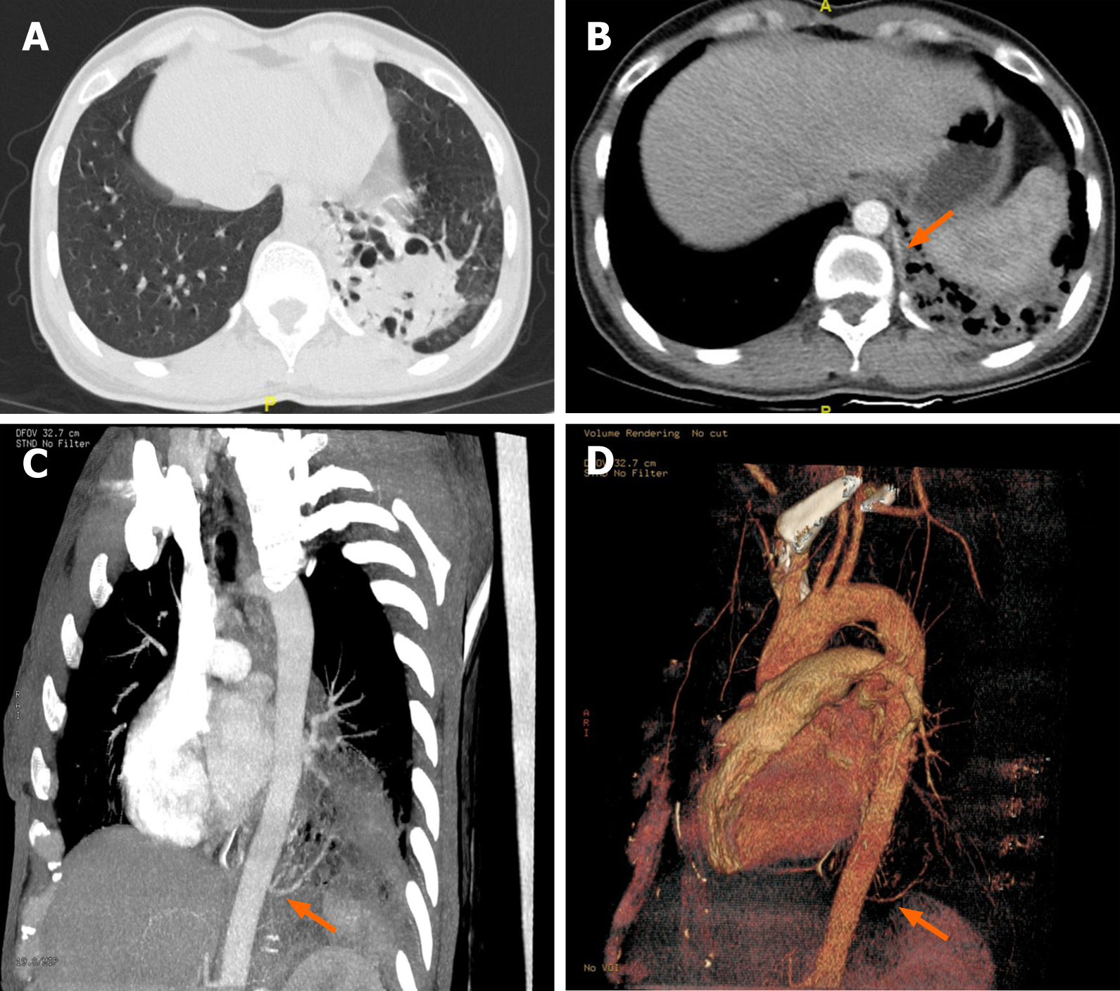
Figure 1 Computed tomography images.
A: High-resolution computed tomography revealed bronchiectasis with honeycomb changes in the lower lobe of the left lung; B: Enhanced scan displayed a vessel from the descending aorta entering the lesion (orange arrow); C: Maximum intensity projection image showed an independent blood vessel originate from the descending aorta (orange arrow); D: Volume rendering image confirmed that the lesions were supplied by an independent vessel (orange arrow).
- Citation: Lin J, Wu XM, Peng MF. Nocardia cyriacigeorgica infection in a patient with pulmonary sequestration: A case report. World J Clin Cases 2021; 9(10): 2367-2372
- URL: https://www.wjgnet.com/2307-8960/full/v9/i10/2367.htm
- DOI: https://dx.doi.org/10.12998/wjcc.v9.i10.2367


