Copyright
©The Author(s) 2021.
World J Clin Cases. Apr 6, 2021; 9(10): 2357-2366
Published online Apr 6, 2021. doi: 10.12998/wjcc.v9.i10.2357
Published online Apr 6, 2021. doi: 10.12998/wjcc.v9.i10.2357
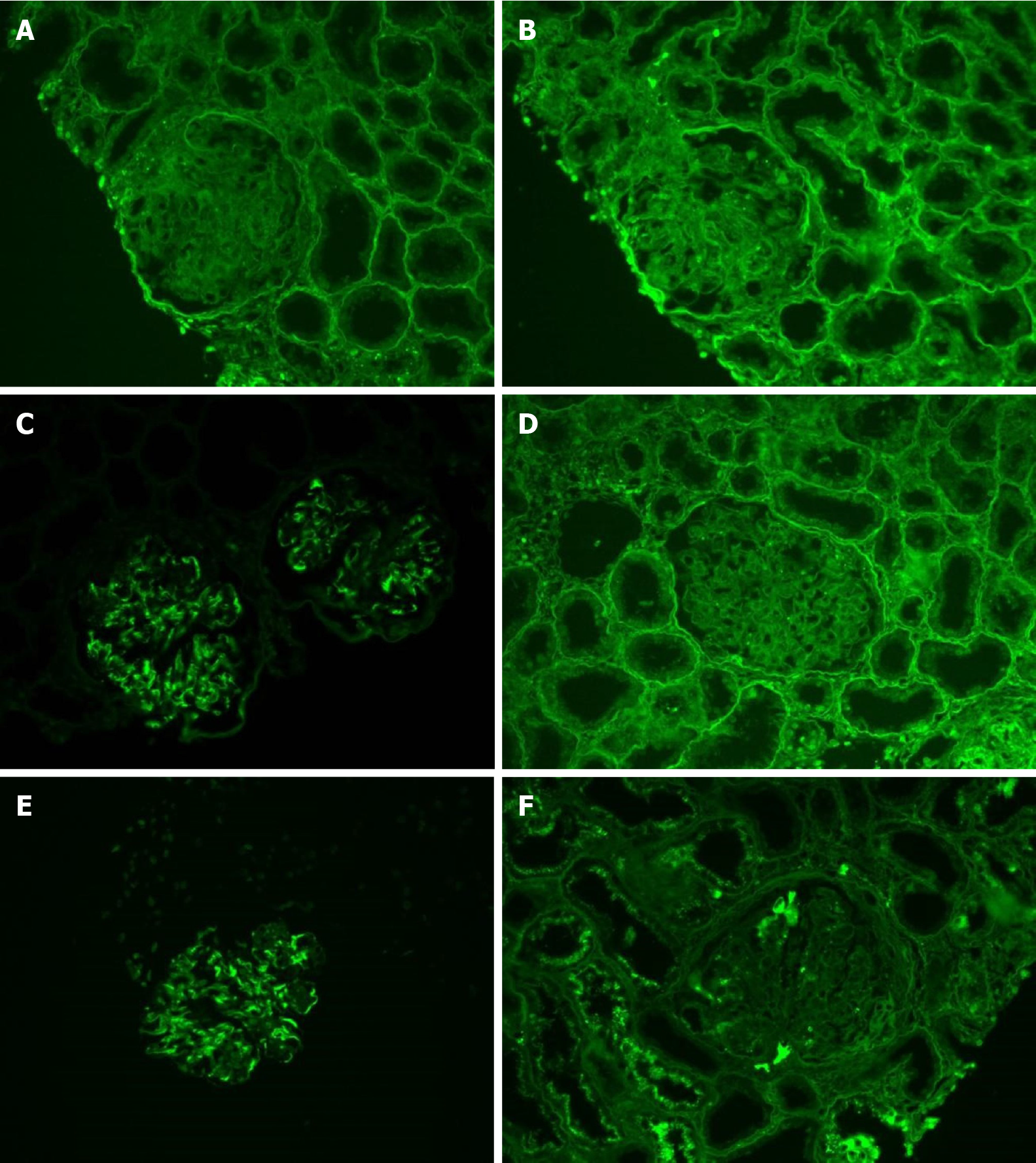
Figure 3 Immunofluorescence analyses.
A-D: Immunofluorescence staining for immunoglobulin G (IgG) subclasses shows intense positivity for (C) IgG3 and negative staining for (A) IgG1, (B) IgG2, and (D) IgG4; E and F: Strong glomerular staining for κ light chain (E) and weak staining for λ light chain (F) were observed. Original magnification, × 400.
- Citation: Xu ZG, Li WL, Wang X, Zhang SY, Zhang YW, Wei X, Li CD, Zeng P, Luan SD. Proliferative glomerulonephritis with monoclonal immunoglobulin G deposits in a young woman: A case report. World J Clin Cases 2021; 9(10): 2357-2366
- URL: https://www.wjgnet.com/2307-8960/full/v9/i10/2357.htm
- DOI: https://dx.doi.org/10.12998/wjcc.v9.i10.2357


