Copyright
©The Author(s) 2021.
World J Clin Cases. Apr 6, 2021; 9(10): 2357-2366
Published online Apr 6, 2021. doi: 10.12998/wjcc.v9.i10.2357
Published online Apr 6, 2021. doi: 10.12998/wjcc.v9.i10.2357
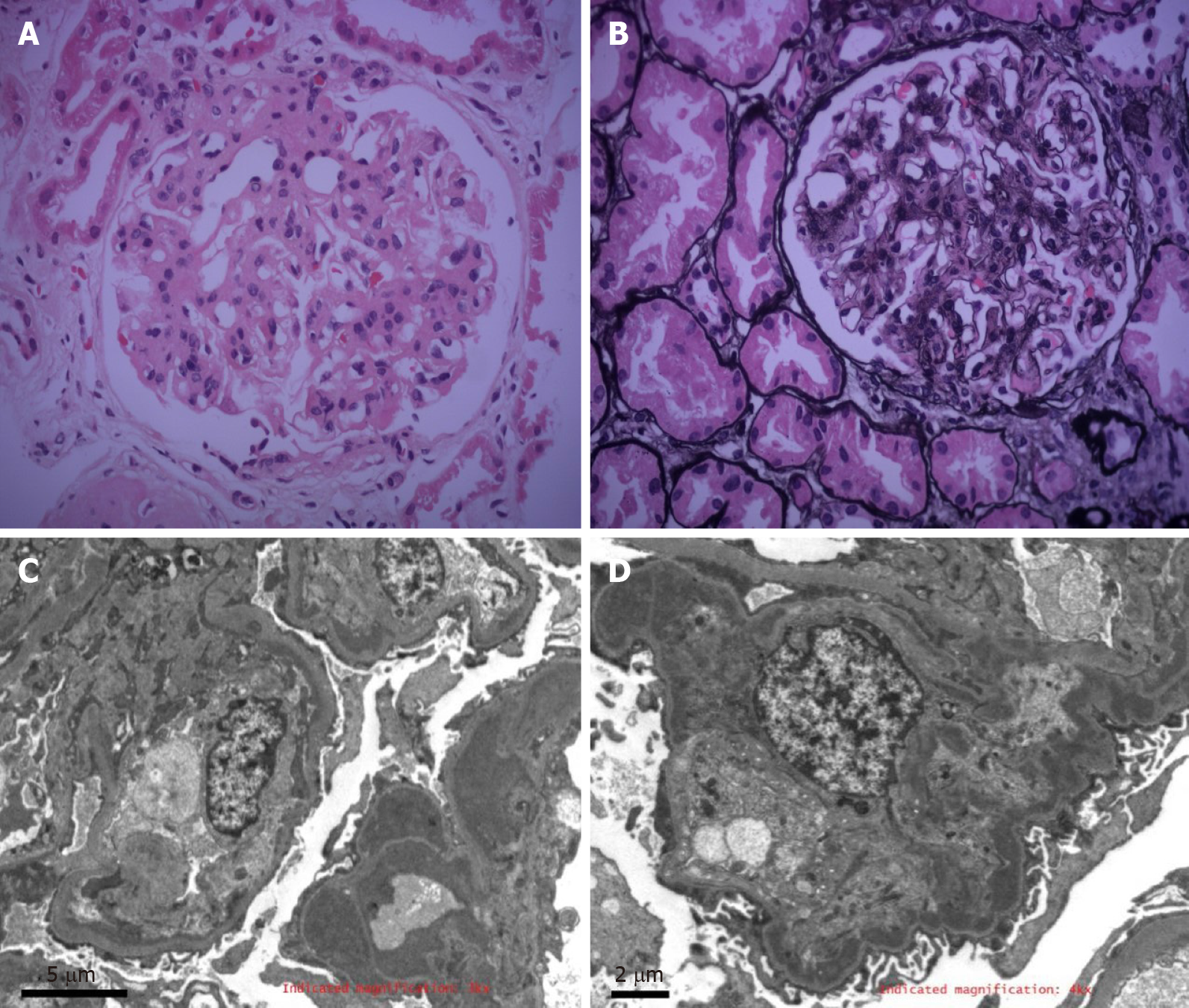
Figure 1 Part of the interstitial region was infiltrated by inflammatory cells.
A and B: Section of the kidney obtained at biopsy showed proliferative lesions of the glomeruli (A: Hematoxylin and eosin staining; B: Periodic acid-Schiff-methenamine silver staining). Original magnification × 400; C and D: Electron-dense deposits were observed in the subendothelial and mesangial areas by electron microscopy.
- Citation: Xu ZG, Li WL, Wang X, Zhang SY, Zhang YW, Wei X, Li CD, Zeng P, Luan SD. Proliferative glomerulonephritis with monoclonal immunoglobulin G deposits in a young woman: A case report. World J Clin Cases 2021; 9(10): 2357-2366
- URL: https://www.wjgnet.com/2307-8960/full/v9/i10/2357.htm
- DOI: https://dx.doi.org/10.12998/wjcc.v9.i10.2357


