Copyright
©The Author(s) 2021.
World J Clin Cases. Apr 6, 2021; 9(10): 2274-2280
Published online Apr 6, 2021. doi: 10.12998/wjcc.v9.i10.2274
Published online Apr 6, 2021. doi: 10.12998/wjcc.v9.i10.2274
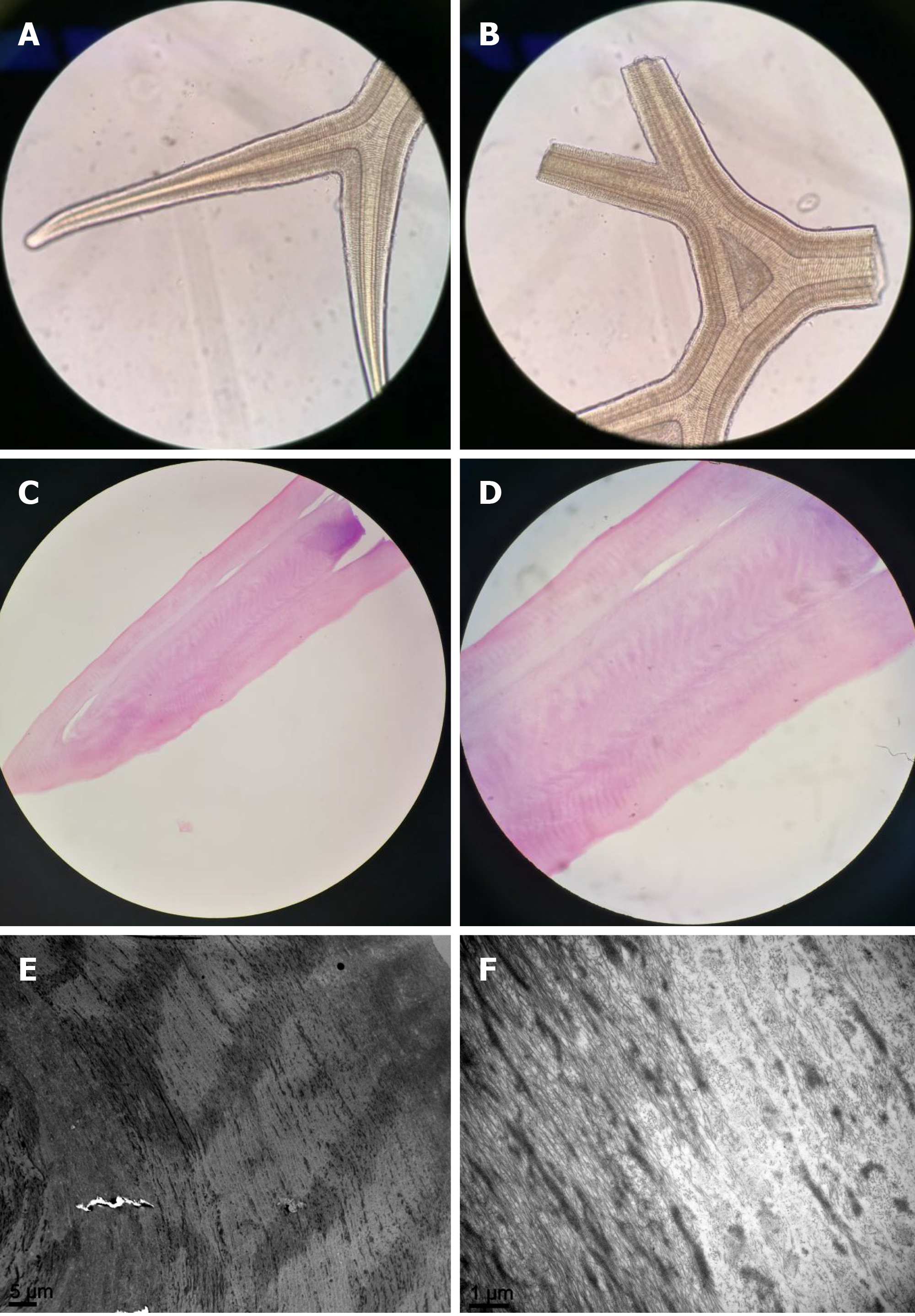
Figure 3 Gross and histologic images.
A and B: Gross microscopic examination showing the scrolls as regular acellular structure like antlers (× 40); C and D: Hematoxylin-eosin staining demonstrated that the scroll comprised eosinophilic acellular tissue (C, × 40; D, × 100); E and F: Scanning electron microscopy showed that the scroll was composed of similar fibrous tissue with a circular arrangement (E, × 2000; F, × 15000).
- Citation: Jin YQ, Hu YP, Dai Q, Wu SQ. Bilateral retrocorneal hyaline scrolls secondary to asymptomatic congenital syphilis: A case report. World J Clin Cases 2021; 9(10): 2274-2280
- URL: https://www.wjgnet.com/2307-8960/full/v9/i10/2274.htm
- DOI: https://dx.doi.org/10.12998/wjcc.v9.i10.2274


