Copyright
©The Author(s) 2021.
World J Clin Cases. Apr 6, 2021; 9(10): 2274-2280
Published online Apr 6, 2021. doi: 10.12998/wjcc.v9.i10.2274
Published online Apr 6, 2021. doi: 10.12998/wjcc.v9.i10.2274
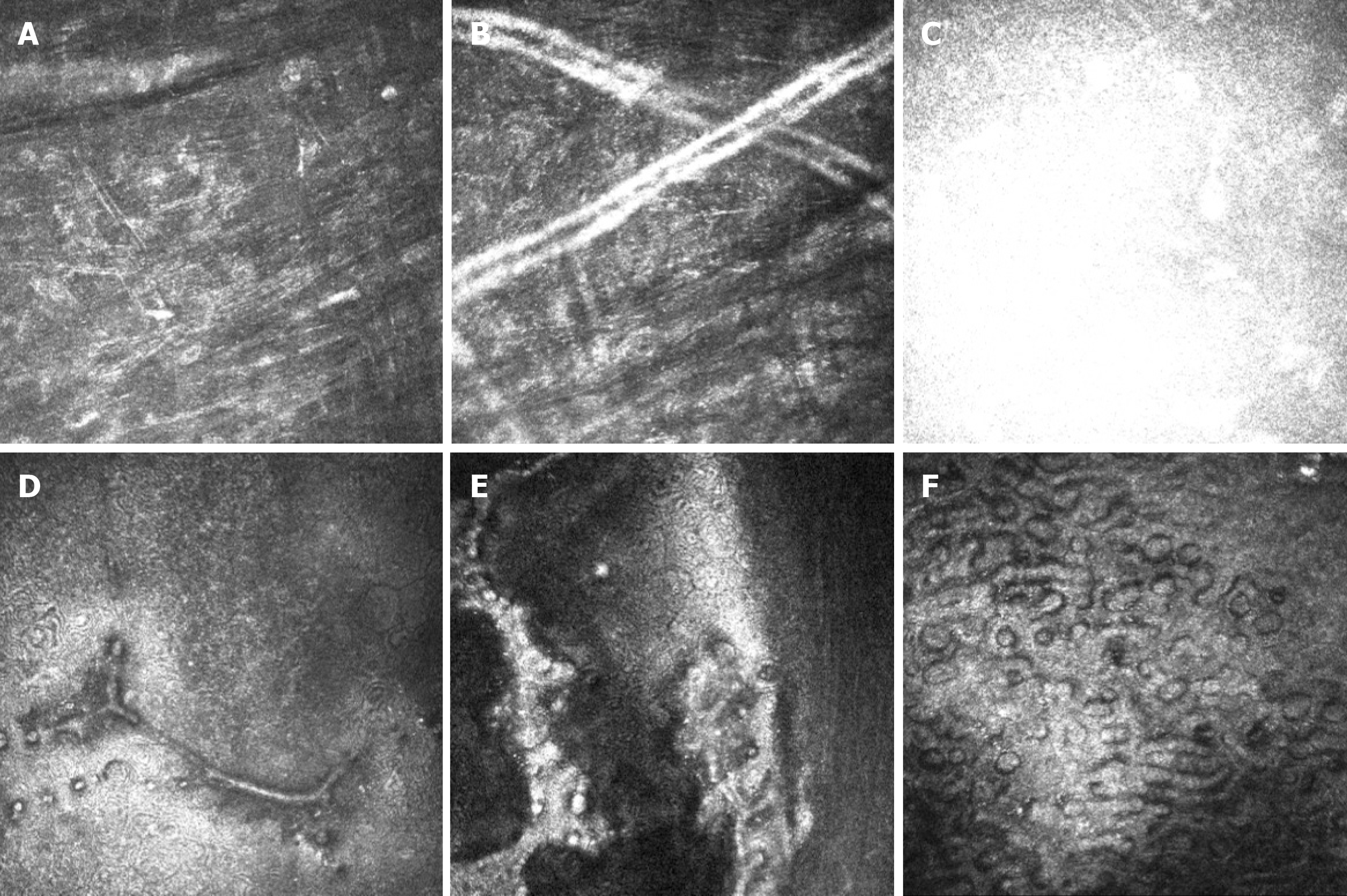
Figure 2 In vivo confocal microscopy showing the changes in the corneas.
A: Activated keratocytes and the altered structure of the extracellular tissue, manifested as thin bright lines in the stroma; B: Tubular structures in the posterior stroma; C: Highly reflective acellular structure at the level of the Descemet’s membrane; D and E: Strip confluent guttae at the level of the corneal endothelium and sheet-like confluent guttae adhesion to the endothelium on the left eye; F: Numerous confluent guttae and enlarged corneal endothelium on the right eye.
- Citation: Jin YQ, Hu YP, Dai Q, Wu SQ. Bilateral retrocorneal hyaline scrolls secondary to asymptomatic congenital syphilis: A case report. World J Clin Cases 2021; 9(10): 2274-2280
- URL: https://www.wjgnet.com/2307-8960/full/v9/i10/2274.htm
- DOI: https://dx.doi.org/10.12998/wjcc.v9.i10.2274


