Copyright
©The Author(s) 2021.
World J Clin Cases. Apr 6, 2021; 9(10): 2274-2280
Published online Apr 6, 2021. doi: 10.12998/wjcc.v9.i10.2274
Published online Apr 6, 2021. doi: 10.12998/wjcc.v9.i10.2274
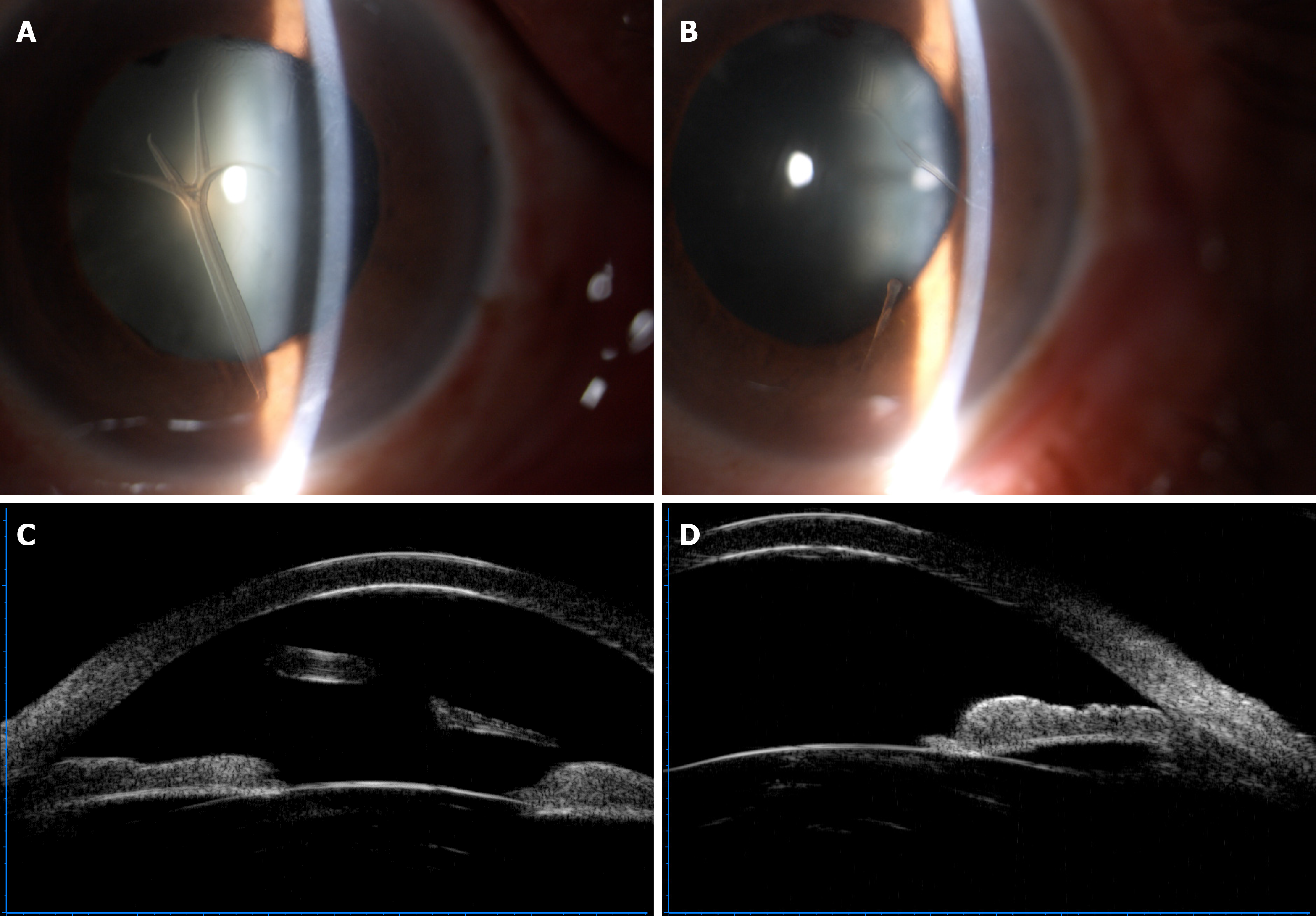
Figure 1 Appearance of retrocorneal scrolls under slit-lamp microscopy and ultrasonic biological microscopy.
A-D: An antler shaped scroll extended to the anterior chamber and attached to the posterior surface of the cornea by a stalk in the right eye (A), and rod-like scrolls adhered to the corneal endothelium in the left eye (B). Free end of the scroll in the left eye (C) and adhered scroll in the right eye (D) with increased reflectivity of the posterior surface of the corneas were observed by ultrasonic biological microscopy.
- Citation: Jin YQ, Hu YP, Dai Q, Wu SQ. Bilateral retrocorneal hyaline scrolls secondary to asymptomatic congenital syphilis: A case report. World J Clin Cases 2021; 9(10): 2274-2280
- URL: https://www.wjgnet.com/2307-8960/full/v9/i10/2274.htm
- DOI: https://dx.doi.org/10.12998/wjcc.v9.i10.2274


