Copyright
©The Author(s) 2021.
World J Clin Cases. Jan 6, 2021; 9(1): 274-277
Published online Jan 6, 2021. doi: 10.12998/wjcc.v9.i1.274
Published online Jan 6, 2021. doi: 10.12998/wjcc.v9.i1.274
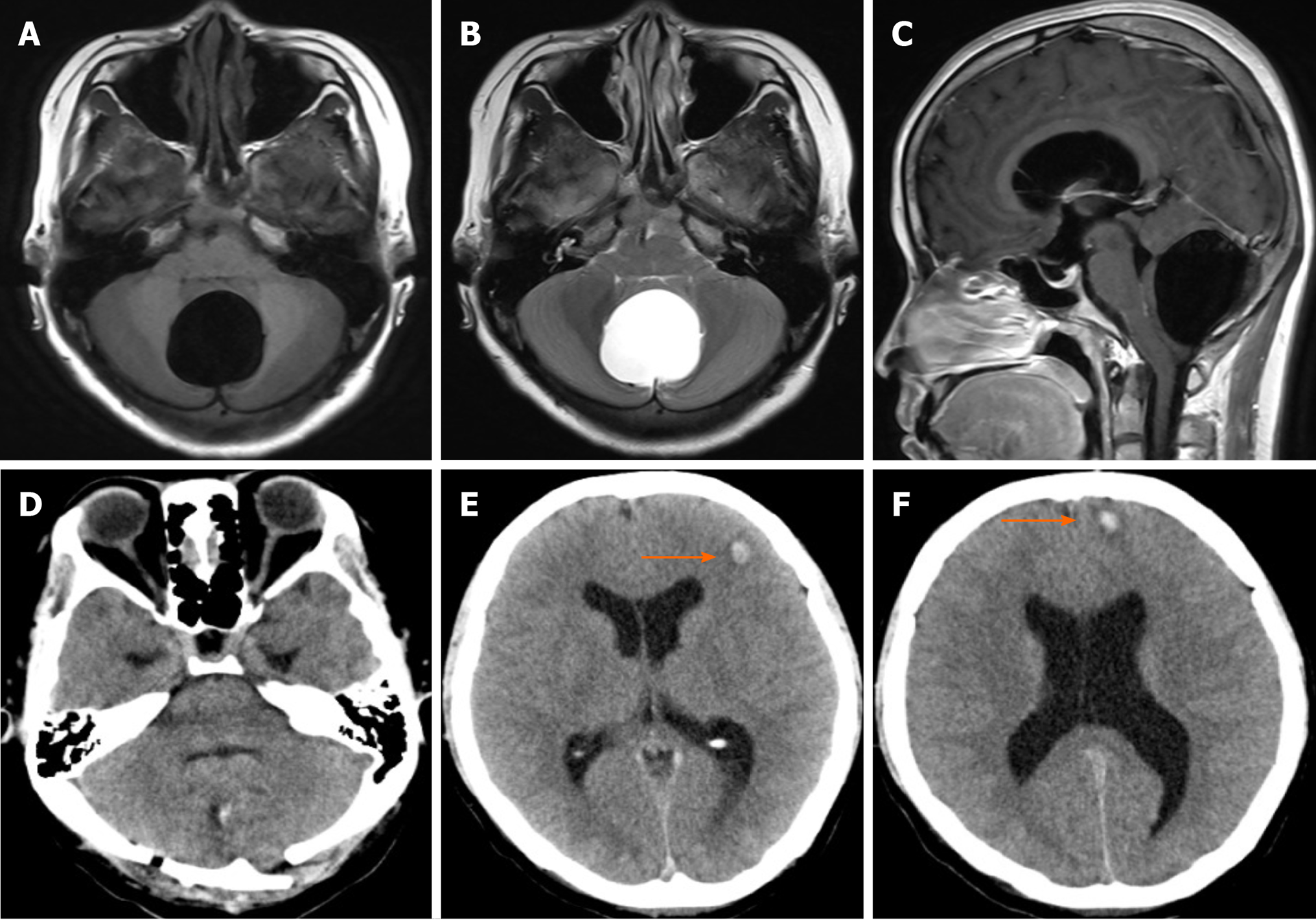
Figure 1 Imaging examinations.
A-C: Axial T1-weighted (A), axial T2-weighted (B) and sagittal T1-weighted (C) magnetic resonance images of the head showing a well-defined lesion of cerebrospinal fluid in the subtentorial region; D: It prompts cysts is good decompression; E and F: Two foci of intraparenchymal hemorrhage were found in the frontal lobe.
- Citation: Wang XJ. Intraparenchymal hemorrhage after surgical decompression of an epencephalon arachnoid cyst: A case report. World J Clin Cases 2021; 9(1): 274-277
- URL: https://www.wjgnet.com/2307-8960/full/v9/i1/274.htm
- DOI: https://dx.doi.org/10.12998/wjcc.v9.i1.274


