Copyright
©The Author(s) 2021.
World J Clin Cases. Jan 6, 2021; 9(1): 211-217
Published online Jan 6, 2021. doi: 10.12998/wjcc.v9.i1.211
Published online Jan 6, 2021. doi: 10.12998/wjcc.v9.i1.211
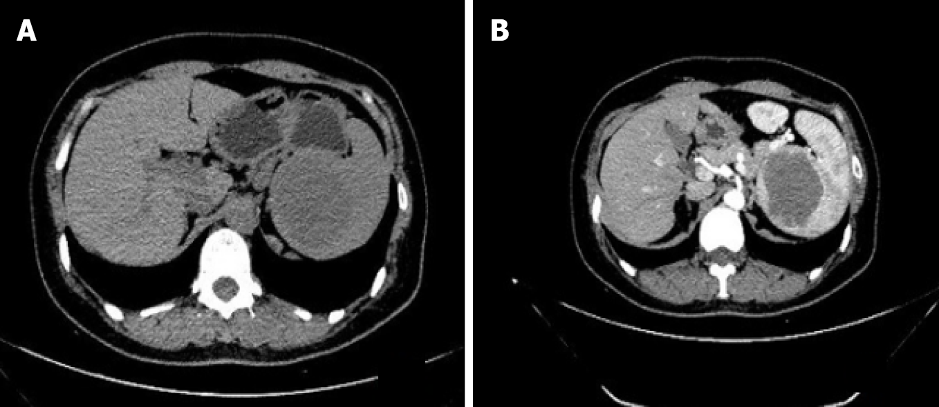
Figure 1 Computed tomography of the abdomen showing the hypodense splenic lesion on non-contrast and contrast-enhanced images.
A: Non-contrast phase of computed tomography showed a hypodense solid lesion, 5.9 cm × 5.1 cm, located at the upper pole of the spleen; B: Contrast-enhanced image showed a mild progressive enhancement of the lesion.
- Citation: Li SX, Fan YH, Wu H, Lv GY. Sclerosing angiomatoid nodular transformation of the spleen: A case report and literature review. World J Clin Cases 2021; 9(1): 211-217
- URL: https://www.wjgnet.com/2307-8960/full/v9/i1/211.htm
- DOI: https://dx.doi.org/10.12998/wjcc.v9.i1.211


