Copyright
©The Author(s) 2021.
World J Clin Cases. Jan 6, 2021; 9(1): 163-169
Published online Jan 6, 2021. doi: 10.12998/wjcc.v9.i1.163
Published online Jan 6, 2021. doi: 10.12998/wjcc.v9.i1.163
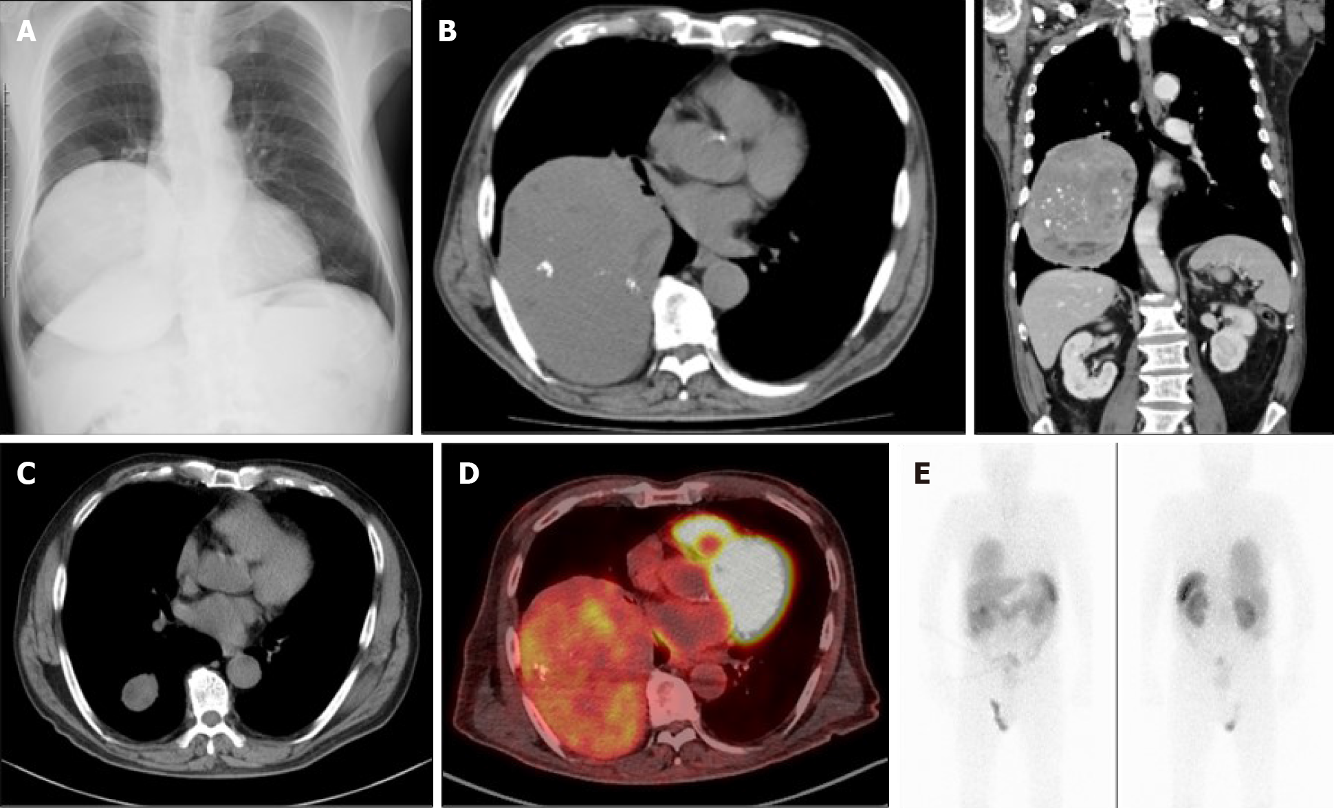
Figure 1 Imaging findings.
A and B: Chest X-ray (A) and computed tomography (B) (left-hand side is transverse plane; right-hand side is coronal plane) showed a heterogenous giant mass measuring 11 cm × 14 cm × 15 cm in size on the right lower chest; C: Computed tomography from ten years ago showed the tumor’s growth in the past decade; D and E: Positron emission tomography (D) and octreotide scintigraphy (E) showed the tumor’s accumulation.
- Citation: Matsumoto S, Yamada E, Nakajima Y, Yamaguchi N, Okamura T, Yajima T, Yoshino S, Horiguchi K, Ishida E, Yoshikawa M, Nagaoka J, Sekiguchi S, Sue M, Okada S, Fukuda I, Shirabe K, Yamada M. Late-onset non-islet cell tumor hypoglycemia: A case report. World J Clin Cases 2021; 9(1): 163-169
- URL: https://www.wjgnet.com/2307-8960/full/v9/i1/163.htm
- DOI: https://dx.doi.org/10.12998/wjcc.v9.i1.163


