Copyright
©The Author(s) 2020.
World J Clin Cases. May 6, 2020; 8(9): 1713-1720
Published online May 6, 2020. doi: 10.12998/wjcc.v8.i9.1713
Published online May 6, 2020. doi: 10.12998/wjcc.v8.i9.1713
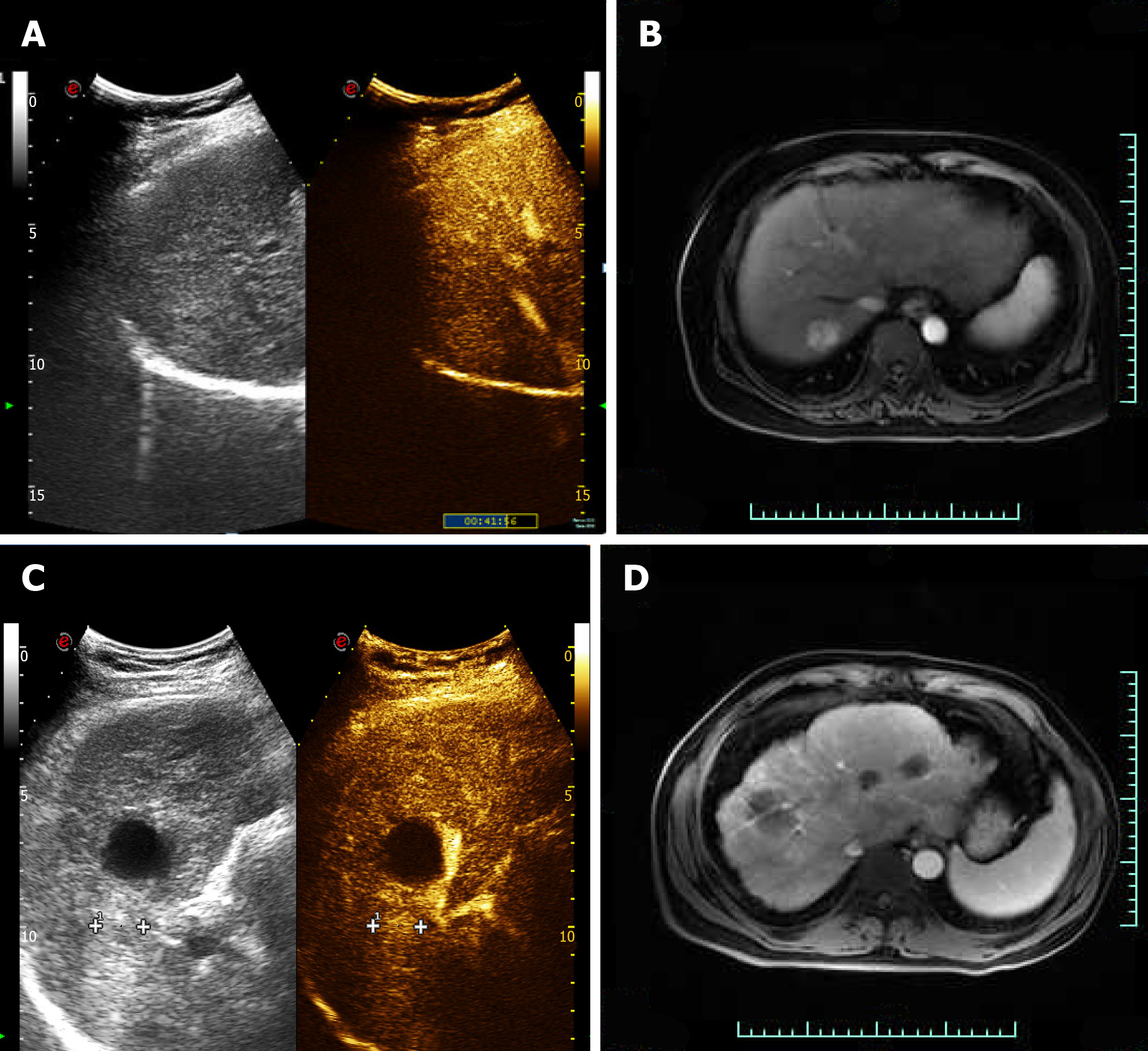
Figure 2 Preoperative contrast-enhanced ultrasound and magnetic resonance imaging findings.
A and B: Case 1. Contrast-enhanced ultrasound of the lesion showed that the lesion was significantly enhanced in the arterial phase (A), and enhanced magnetic resonance imaging detected a round long T1 and long T2 signal in the upper posterior segment of the right liver lobe near the liver, with the lesion significantly enhanced in the arterial phase; C and D: Case 2. Contrast-enhanced ultrasound (C) enhanced magnetic resonance imaging (D) showed high enhancement in the arterial phase.
- Citation: Cao N, Cai HJ, Sun XX, Liu DL, Huang B. Application of curved ablation in liver cancer with special morphology or location: Report of two cases. World J Clin Cases 2020; 8(9): 1713-1720
- URL: https://www.wjgnet.com/2307-8960/full/v8/i9/1713.htm
- DOI: https://dx.doi.org/10.12998/wjcc.v8.i9.1713


