Copyright
©The Author(s) 2020.
World J Clin Cases. Apr 26, 2020; 8(8): 1502-1506
Published online Apr 26, 2020. doi: 10.12998/wjcc.v8.i8.1502
Published online Apr 26, 2020. doi: 10.12998/wjcc.v8.i8.1502
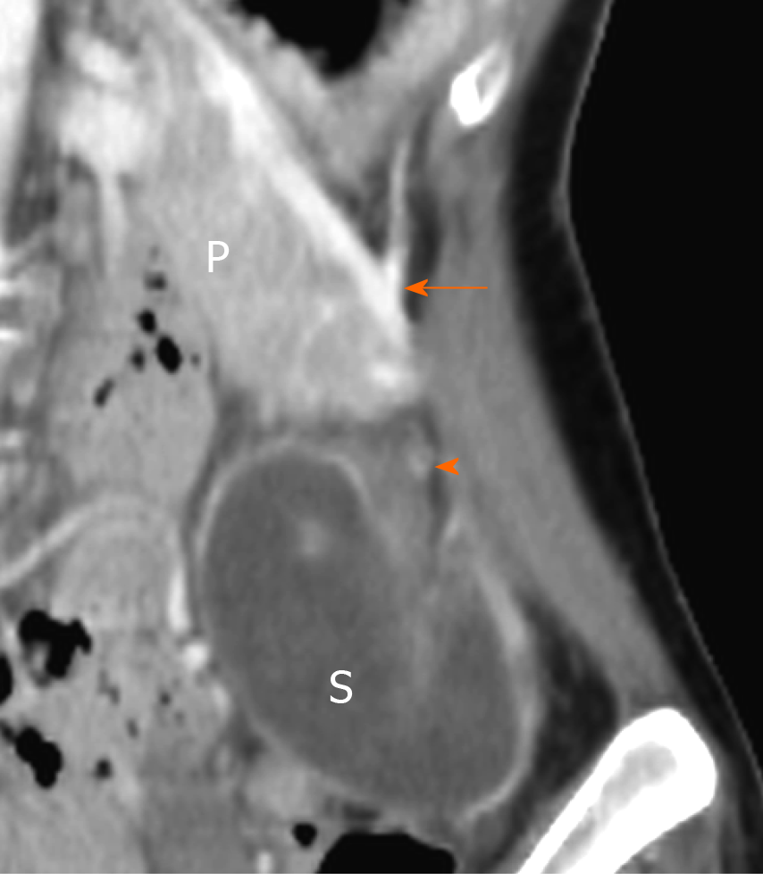
Figure 1 Contrast enhanced computed tomography showed a non-homogeneously enhanced splenic parenchyma below the pancreatic tail in the left iliac fossa.
P: Pancreas; S: Spleen; Arrow: Splenic vessel; Arrowhead: Unenhanced splenic hilum.
- Citation: Chang YL, Lin J, Li YH, Tsao LC. Unusual association of Axenfeld-Rieger syndrome and wandering spleen: A case report. World J Clin Cases 2020; 8(8): 1502-1506
- URL: https://www.wjgnet.com/2307-8960/full/v8/i8/1502.htm
- DOI: https://dx.doi.org/10.12998/wjcc.v8.i8.1502


