Copyright
©The Author(s) 2020.
World J Clin Cases. Apr 6, 2020; 8(7): 1319-1325
Published online Apr 6, 2020. doi: 10.12998/wjcc.v8.i7.1319
Published online Apr 6, 2020. doi: 10.12998/wjcc.v8.i7.1319
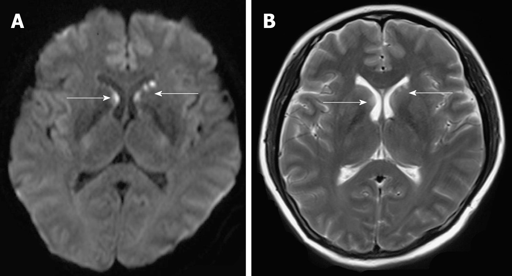
Figure 1 Brain magnetic resonance imaging.
A: Diffusion-weighted imaging showed high signals (arrow) in the areas beside the frontal horn of the lateral ventricle; B: T2 weight imaging revealed slightly higher signal (arrow) in the areas beside the frontal horn of the lateral ventricle.
- Citation: Li JA, Cheng YY, Cui ZT, Jiang W, Zhang WQ, Du ZH, Gao B, Xie YY, Meng HM. Disseminated histoplasmosis in primary Sjögren syndrome: A case report. World J Clin Cases 2020; 8(7): 1319-1325
- URL: https://www.wjgnet.com/2307-8960/full/v8/i7/1319.htm
- DOI: https://dx.doi.org/10.12998/wjcc.v8.i7.1319


