Copyright
©The Author(s) 2020.
World J Clin Cases. Apr 6, 2020; 8(7): 1287-1294
Published online Apr 6, 2020. doi: 10.12998/wjcc.v8.i7.1287
Published online Apr 6, 2020. doi: 10.12998/wjcc.v8.i7.1287
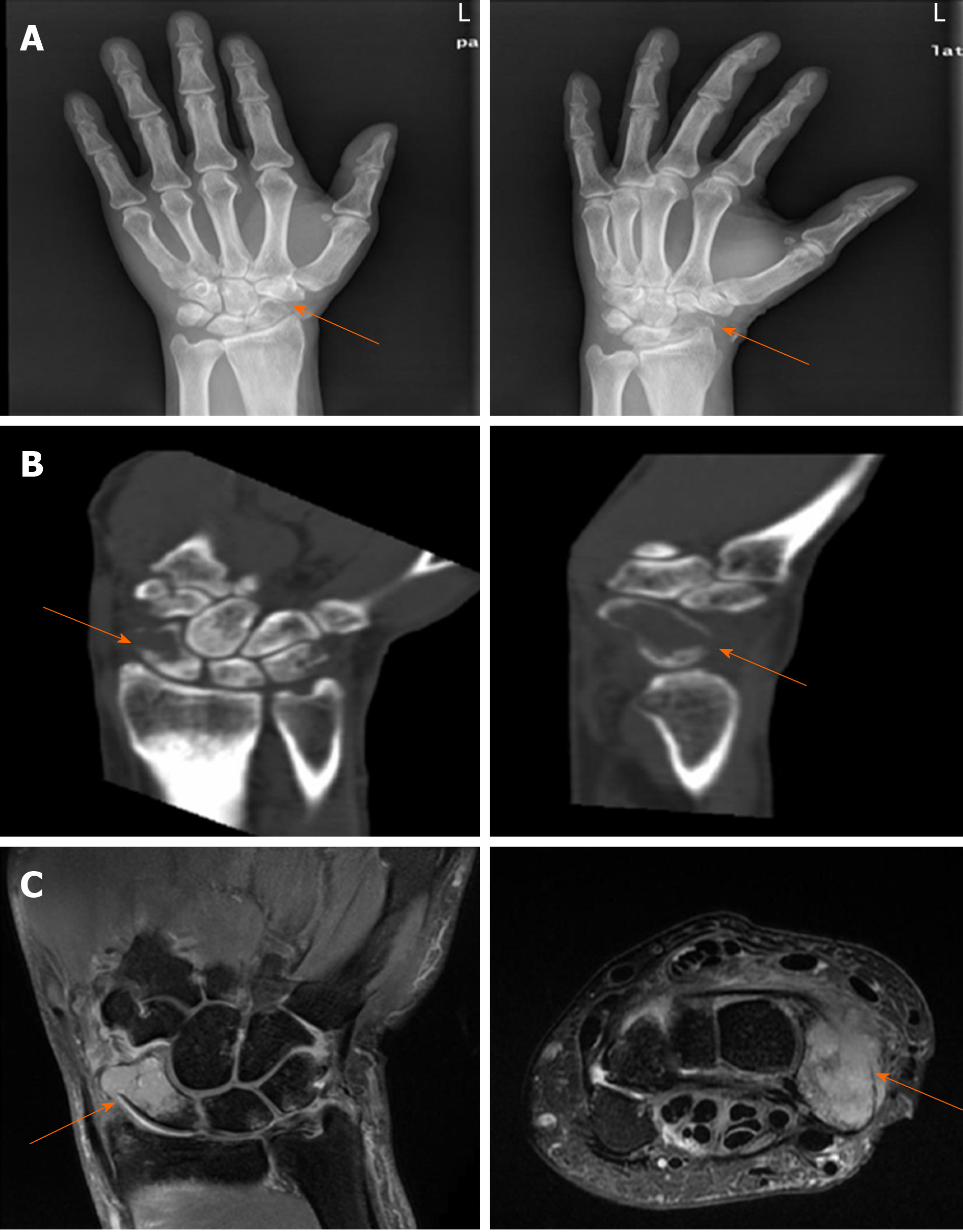
Figure 1 Imaging findings in the left hand.
A: Plain X-ray showed a decrease in scaphoid bone density accompanied by a fracture; B and C: Computed tomography and magnetic resonance imaging scans demonstrated scaphoid and triangular bone destruction and soft tissue swelling, indicating a pathological fracture.
- Citation: Zhang YJ, Wang YY, Yang Q, Li JB. Scaphoid metastasis as the first sign of occult gastroesophageal junction cancer: A case report. World J Clin Cases 2020; 8(7): 1287-1294
- URL: https://www.wjgnet.com/2307-8960/full/v8/i7/1287.htm
- DOI: https://dx.doi.org/10.12998/wjcc.v8.i7.1287


