Copyright
©The Author(s) 2020.
World J Clin Cases. Apr 6, 2020; 8(7): 1278-1286
Published online Apr 6, 2020. doi: 10.12998/wjcc.v8.i7.1278
Published online Apr 6, 2020. doi: 10.12998/wjcc.v8.i7.1278
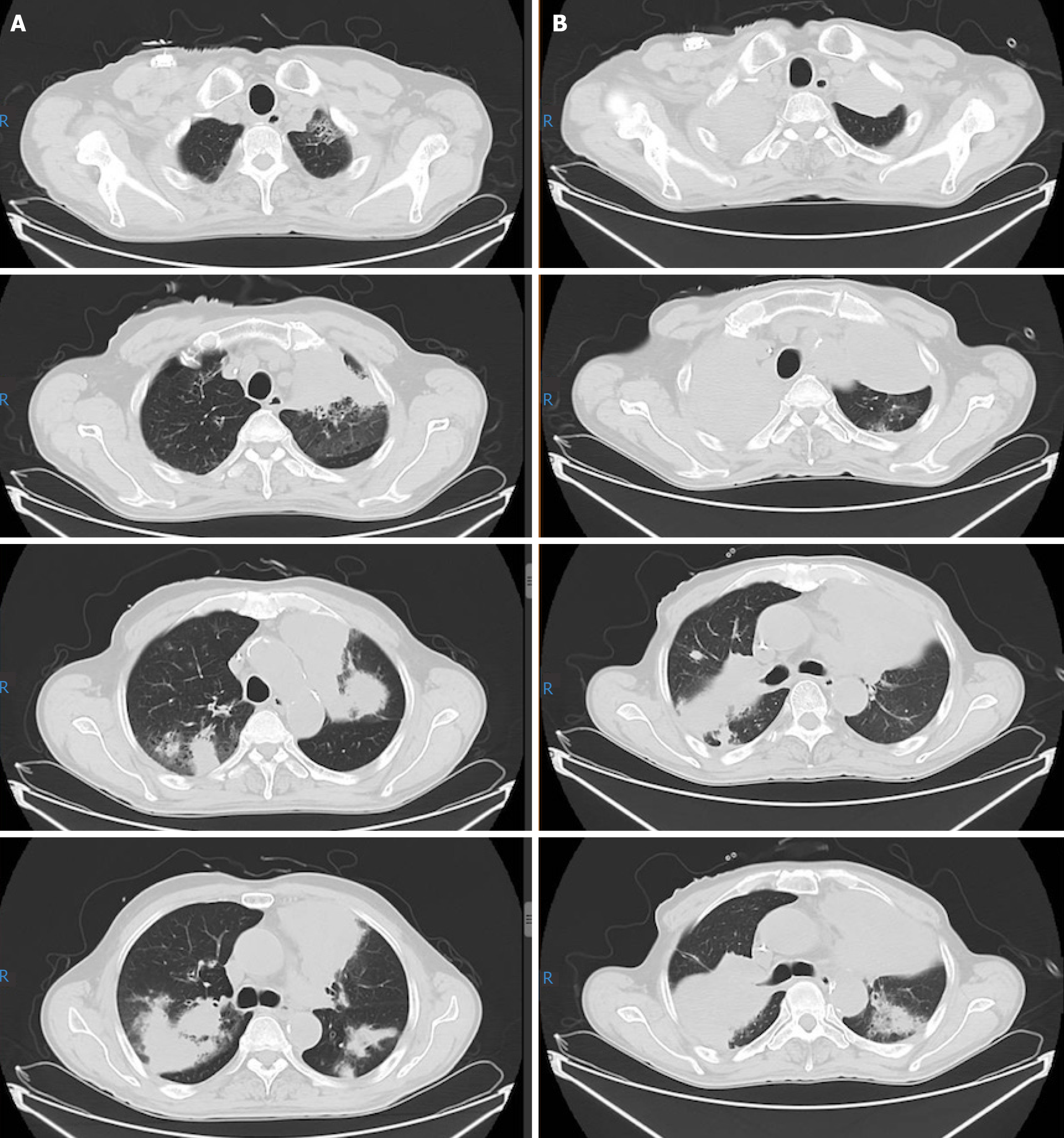
Figure 3 Thoracic computed tomography scan of case one taken at recurrence 11 mo following his diagnosis and re-evaluation after three cycles of rescue chemotherapy.
A: Computed tomography taken before rescue therapy. It revealed multiple lesions in the bilateral lungs; B: Computed tomography taken after rescue therapy. It showed that the shadow in bilateral lungs aggravated.
- Citation: Liu TZ, Zheng YJ, Zhang ZW, Li SS, Chen JT, Peng AH, Huang RW. Chidamide based combination regimen for treatment of monomorphic epitheliotropic intestinal T cell lymphoma following radical operation: Two case reports. World J Clin Cases 2020; 8(7): 1278-1286
- URL: https://www.wjgnet.com/2307-8960/full/v8/i7/1278.htm
- DOI: https://dx.doi.org/10.12998/wjcc.v8.i7.1278


