Copyright
©The Author(s) 2020.
World J Clin Cases. Apr 6, 2020; 8(7): 1271-1277
Published online Apr 6, 2020. doi: 10.12998/wjcc.v8.i7.1271
Published online Apr 6, 2020. doi: 10.12998/wjcc.v8.i7.1271
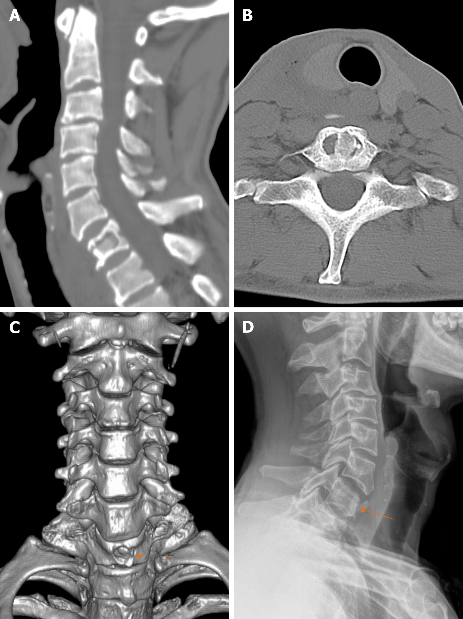
Figure 3 Imaging findings after treatment.
A-C: The axial computed tomography (CT) images (A), sagittal CT images (B) and the Three-dimensional CT reconstruction (C) of the cervical vertebra after the operation showed a good position for bone grafting; D: The last follow-up cervical X-ray showed no radiological recurrence.
- Citation: Xu ZQ, Zhang P, Zhong ZH, Zhou W, Yu HT. Spinal intraosseous schwannoma without spinal canal and neuroforamina involvement: A case report. World J Clin Cases 2020; 8(7): 1271-1277
- URL: https://www.wjgnet.com/2307-8960/full/v8/i7/1271.htm
- DOI: https://dx.doi.org/10.12998/wjcc.v8.i7.1271


