Copyright
©The Author(s) 2020.
World J Clin Cases. Apr 6, 2020; 8(7): 1203-1212
Published online Apr 6, 2020. doi: 10.12998/wjcc.v8.i7.1203
Published online Apr 6, 2020. doi: 10.12998/wjcc.v8.i7.1203
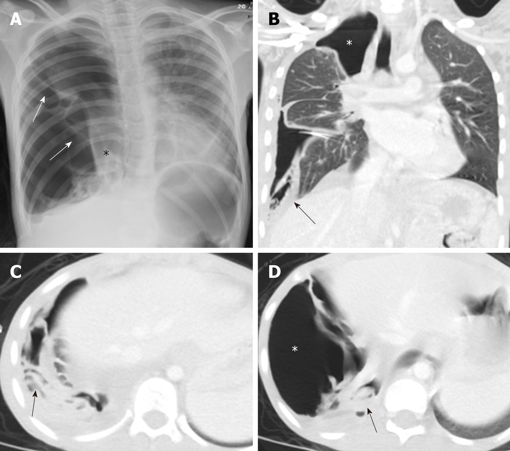
Figure 6 A 10-year old female with spontaneous ruptured lung hydatid cyst presenting with pneumothorax and complicated by broncho-pleural fistula.
Patient presented with oxygen desaturation and chest pain with chronic history of shortness of breath and cough for 3 months. A: Frontal chest radiograph shows large right-sided pneumothorax with small pleural effusion and multiple pleural septations (arrows), medially displaced collapsed right lung (asterisk), mild contralateral shift of the mediastinum to the left, and flattening of the right hemidiaphragm; B-D: Coronal and axial images of chest computed tomography scan after placement of chest tube show improvement of lung aeration with persistent large right-sided pneumothorax (asterisks) which suggest broncho-plural fistula, multiple collapsed membranes (arrows), and adjacent plural effusion.
- Citation: Saeedan MB, Aljohani IM, Alghofaily KA, Loutfi S, Ghosh S. Thoracic hydatid disease: A radiologic review of unusual cases. World J Clin Cases 2020; 8(7): 1203-1212
- URL: https://www.wjgnet.com/2307-8960/full/v8/i7/1203.htm
- DOI: https://dx.doi.org/10.12998/wjcc.v8.i7.1203


