Copyright
©The Author(s) 2020.
World J Clin Cases. Apr 6, 2020; 8(7): 1203-1212
Published online Apr 6, 2020. doi: 10.12998/wjcc.v8.i7.1203
Published online Apr 6, 2020. doi: 10.12998/wjcc.v8.i7.1203
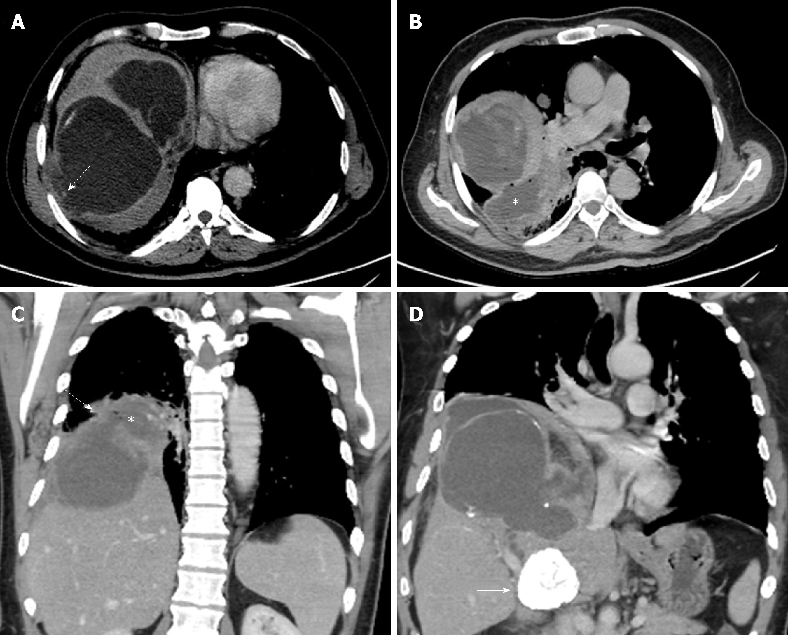
Figure 5 A 42-year old male presented with liver hydatid cyst complicated by broncho-biliary fistula presented with coughing bile.
Axial and coronal computed tomography scan images using liver window (A) and standard soft tissue window (B-D) show intercommunicating multiple liver cystic lesions with internal non-enhancing septations and peripheral calcifications within hepatic segments 8 and 7. The right lower lobe lung cystic lesion (asterisks) contains multiple air foci indicating bronchial communication with evidence of wide communication with the hepatic cystic lesion through the diaphragmatic defect (dashed arrows in A and C). A calcified hepatic lesion near the porta hepatis represents a healed hydatid cyst (arrow in D).
- Citation: Saeedan MB, Aljohani IM, Alghofaily KA, Loutfi S, Ghosh S. Thoracic hydatid disease: A radiologic review of unusual cases. World J Clin Cases 2020; 8(7): 1203-1212
- URL: https://www.wjgnet.com/2307-8960/full/v8/i7/1203.htm
- DOI: https://dx.doi.org/10.12998/wjcc.v8.i7.1203


