Copyright
©The Author(s) 2020.
World J Clin Cases. Apr 6, 2020; 8(7): 1203-1212
Published online Apr 6, 2020. doi: 10.12998/wjcc.v8.i7.1203
Published online Apr 6, 2020. doi: 10.12998/wjcc.v8.i7.1203
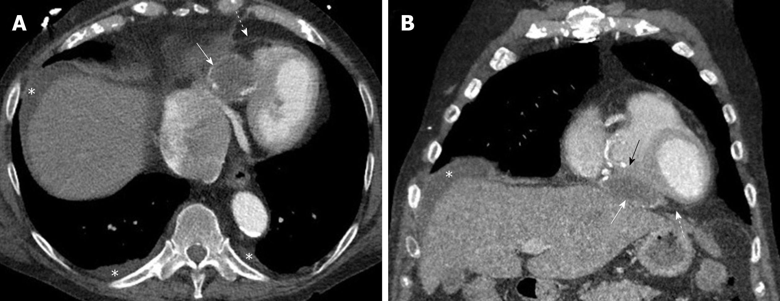
Figure 3 Pericardial hydatid cyst.
A, B: Axial and coronal contrast enhanced computed tomography images show a partially peripherally calcified cystic lesion (white arrows) along the inferior aspect of the heart and within the pericardial sac (dashed arrows) with mild mass effect and displacement of the right ventricle (black arrow). Small pleural effusions and ascites are shown (asterisks).
- Citation: Saeedan MB, Aljohani IM, Alghofaily KA, Loutfi S, Ghosh S. Thoracic hydatid disease: A radiologic review of unusual cases. World J Clin Cases 2020; 8(7): 1203-1212
- URL: https://www.wjgnet.com/2307-8960/full/v8/i7/1203.htm
- DOI: https://dx.doi.org/10.12998/wjcc.v8.i7.1203


