Copyright
©The Author(s) 2020.
World J Clin Cases. Apr 6, 2020; 8(7): 1203-1212
Published online Apr 6, 2020. doi: 10.12998/wjcc.v8.i7.1203
Published online Apr 6, 2020. doi: 10.12998/wjcc.v8.i7.1203
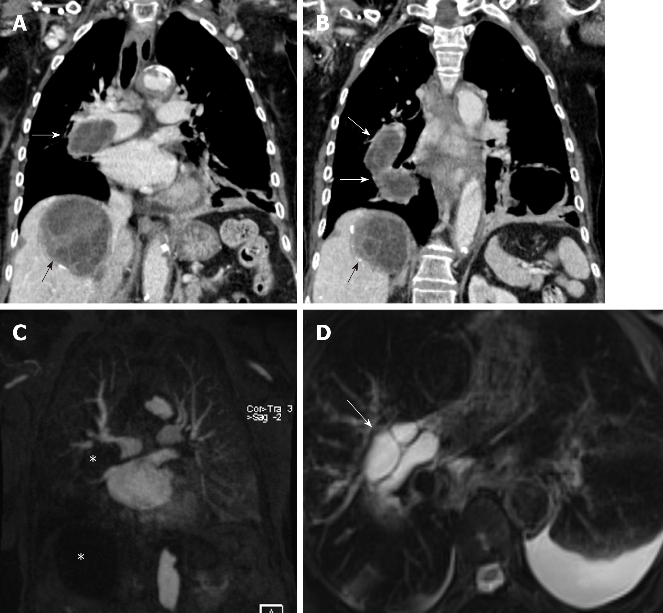
Figure 2 An 82-year-old female with pulmonary artery hydatid cyst embolism presented with shortness of breath.
A, B: Coronal images of enhanced computed tomography scan show right main pulmonary artery intra-luminal cystic filling defects causing expansion and near occlusion of the artery with extension into the right interlobar pulmonary artery (white arrows). There is hepatic hydatid cyst with internal septation and daughter cysts and scattered wall calcifications (black arrows); C: Coronal image of T1 weighted magnetic resonance angiography confirms the presence of filling defect of low signal intensity within the right pulmonary artery and low signal intensity liver lesion (asterisks) with no evidence of contrast enhancement; D: Axial T2 weighted image with fat saturation shows right main pulmonary artery intra-luminal cystic filling defects of high signal intensity with septations suggestive of daughter cysts (arrow). Left pleural effusion is noted.
- Citation: Saeedan MB, Aljohani IM, Alghofaily KA, Loutfi S, Ghosh S. Thoracic hydatid disease: A radiologic review of unusual cases. World J Clin Cases 2020; 8(7): 1203-1212
- URL: https://www.wjgnet.com/2307-8960/full/v8/i7/1203.htm
- DOI: https://dx.doi.org/10.12998/wjcc.v8.i7.1203


