Copyright
©The Author(s) 2020.
World J Clin Cases. Apr 6, 2020; 8(7): 1203-1212
Published online Apr 6, 2020. doi: 10.12998/wjcc.v8.i7.1203
Published online Apr 6, 2020. doi: 10.12998/wjcc.v8.i7.1203
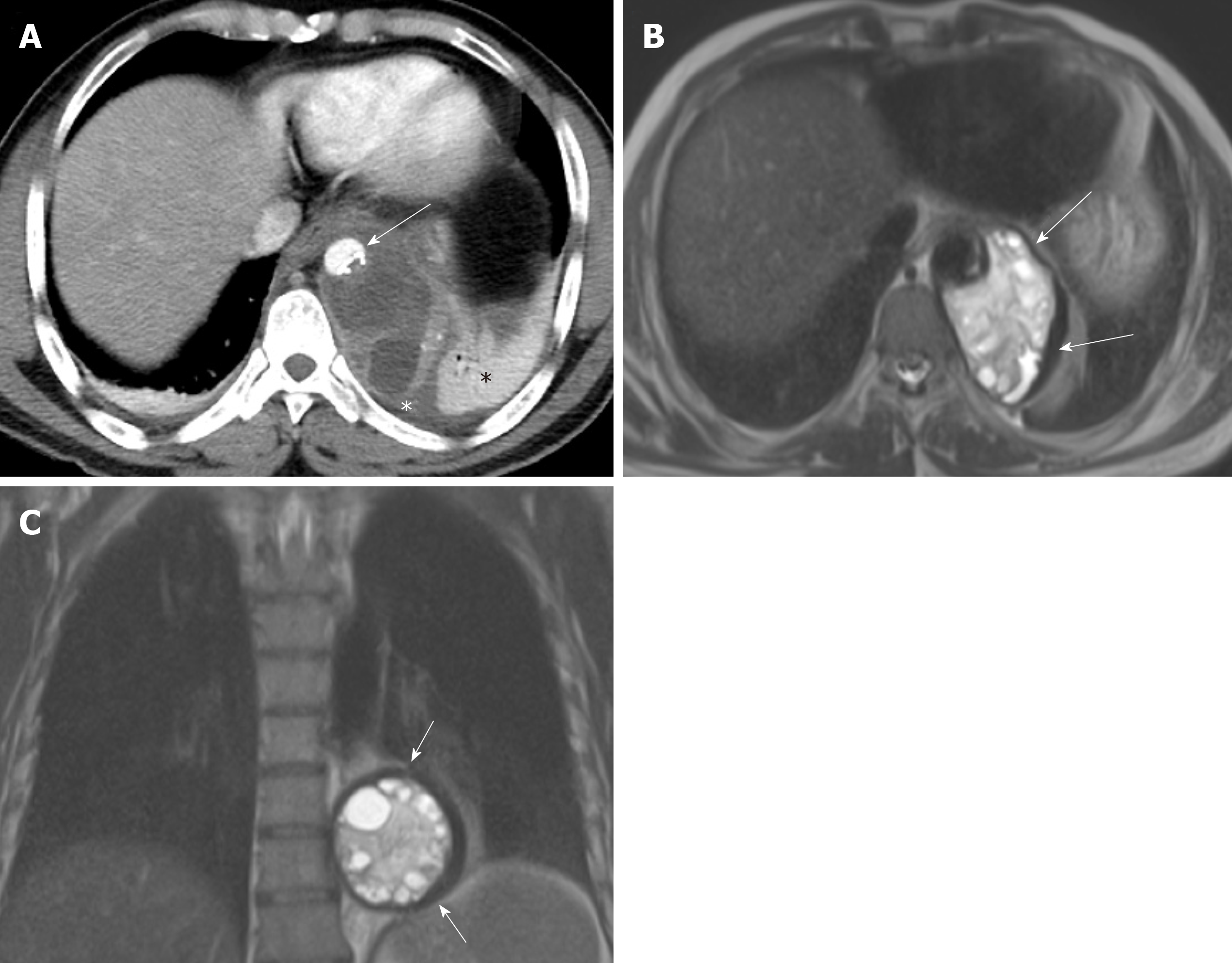
Figure 1 A 48-year old male with mediastinal hydatid cyst presented with acute chest pain.
A: Axial image of enhanced computed tomography angiogram shows a complex cystic lesion with thick wall (white arrows) that is predominately located in the left side of the posterior mediastinum with evidence small calcifications at the periphery of the lesions at the site of abutment with descending thoracic aorta. There is adjacent pleural effusion (white asterisk) and enhancing left lower lobe atelectasis (white asterisk); B, C: Axial and coronal T2 weighted images show a complex cystic lesion with thick dark rim (arrows) abutting the descending thoracic aorta with multiple peripheral daughter cysts of higher signal intensity compared to the intermediately hyperintenese matrix of the mother cyst. Magnetic resonance imaging findings are highly suggestive of hydatid cyst, likely primary given absence of evidence of lung and liver hydatid disease.
- Citation: Saeedan MB, Aljohani IM, Alghofaily KA, Loutfi S, Ghosh S. Thoracic hydatid disease: A radiologic review of unusual cases. World J Clin Cases 2020; 8(7): 1203-1212
- URL: https://www.wjgnet.com/2307-8960/full/v8/i7/1203.htm
- DOI: https://dx.doi.org/10.12998/wjcc.v8.i7.1203


