Copyright
©The Author(s) 2020.
World J Clin Cases. Mar 26, 2020; 8(6): 1164-1171
Published online Mar 26, 2020. doi: 10.12998/wjcc.v8.i6.1164
Published online Mar 26, 2020. doi: 10.12998/wjcc.v8.i6.1164
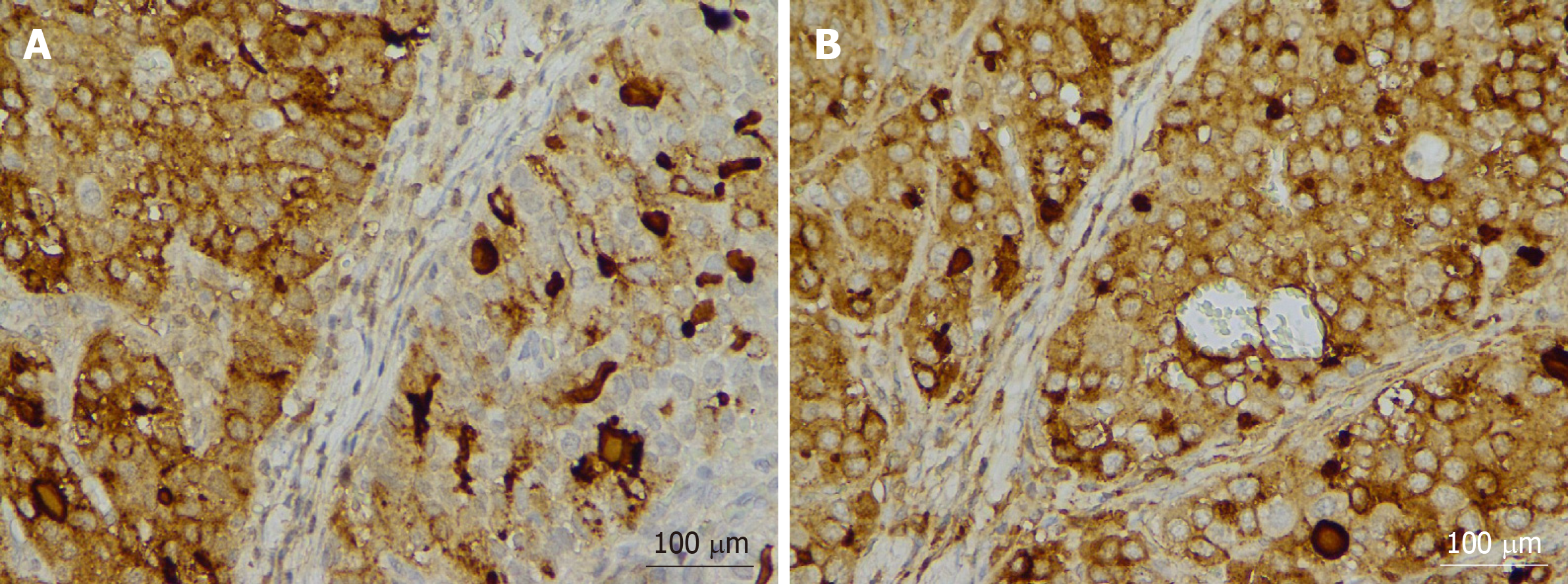
Figure 2 Light microscopy images of a lesion stained immunohistochemically.
A: Hepatocyte+; B: Alpha-fetoprotein+. Original magnification, × 400.
- Citation: Zhang ZR, Wu J, Li HW, Wang T. Hepatoid adenocarcinoma of the stomach: Thirteen case reports and review of literature. World J Clin Cases 2020; 8(6): 1164-1171
- URL: https://www.wjgnet.com/2307-8960/full/v8/i6/1164.htm
- DOI: https://dx.doi.org/10.12998/wjcc.v8.i6.1164


