Copyright
©The Author(s) 2020.
World J Clin Cases. Mar 26, 2020; 8(6): 1158-1163
Published online Mar 26, 2020. doi: 10.12998/wjcc.v8.i6.1158
Published online Mar 26, 2020. doi: 10.12998/wjcc.v8.i6.1158
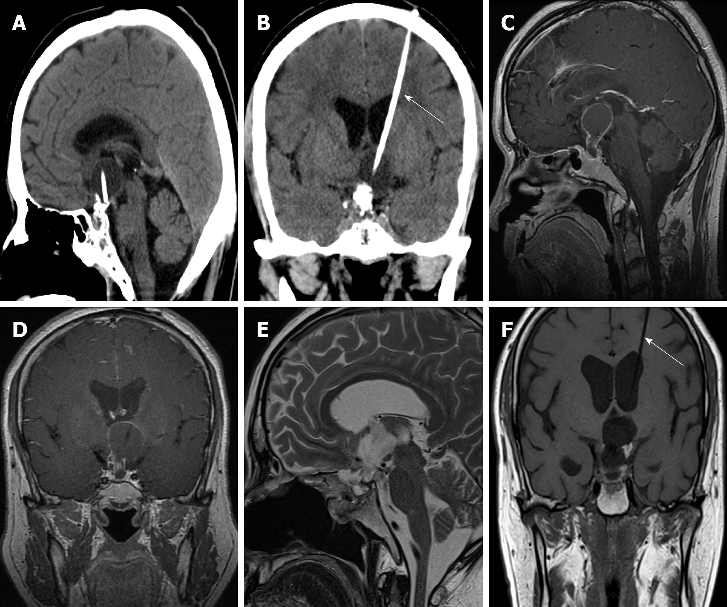
Figure 1 Imaging findings.
A and B: Head computed tomography before transsphenoidal surgery showed calcified cystic mass components in the intra- and suprasellar region, and internal irradiation tube in the cystic cavity of the mass (white arrow); C and D: Magnetic resonance imaging before transsphenoidal surgery showed a 2.7 cm × 2.5 cm × 2.5 cm calcified cystic intra- and suprasellar mass, which was strongly enhanced after injection of contrast medium; E and F: Magnetic resonance imaging after transsphenoidal surgery showed complete tumor resection and irradiation tube was reserved (white arrow).
- Citation: Chang T, Yang YL, Gao L, Li LH. Cerebral venous sinus thrombosis following transsphenoidal surgery for craniopharyngioma: A case report. World J Clin Cases 2020; 8(6): 1158-1163
- URL: https://www.wjgnet.com/2307-8960/full/v8/i6/1158.htm
- DOI: https://dx.doi.org/10.12998/wjcc.v8.i6.1158


