Copyright
©The Author(s) 2020.
World J Clin Cases. Mar 26, 2020; 8(6): 1129-1136
Published online Mar 26, 2020. doi: 10.12998/wjcc.v8.i6.1129
Published online Mar 26, 2020. doi: 10.12998/wjcc.v8.i6.1129
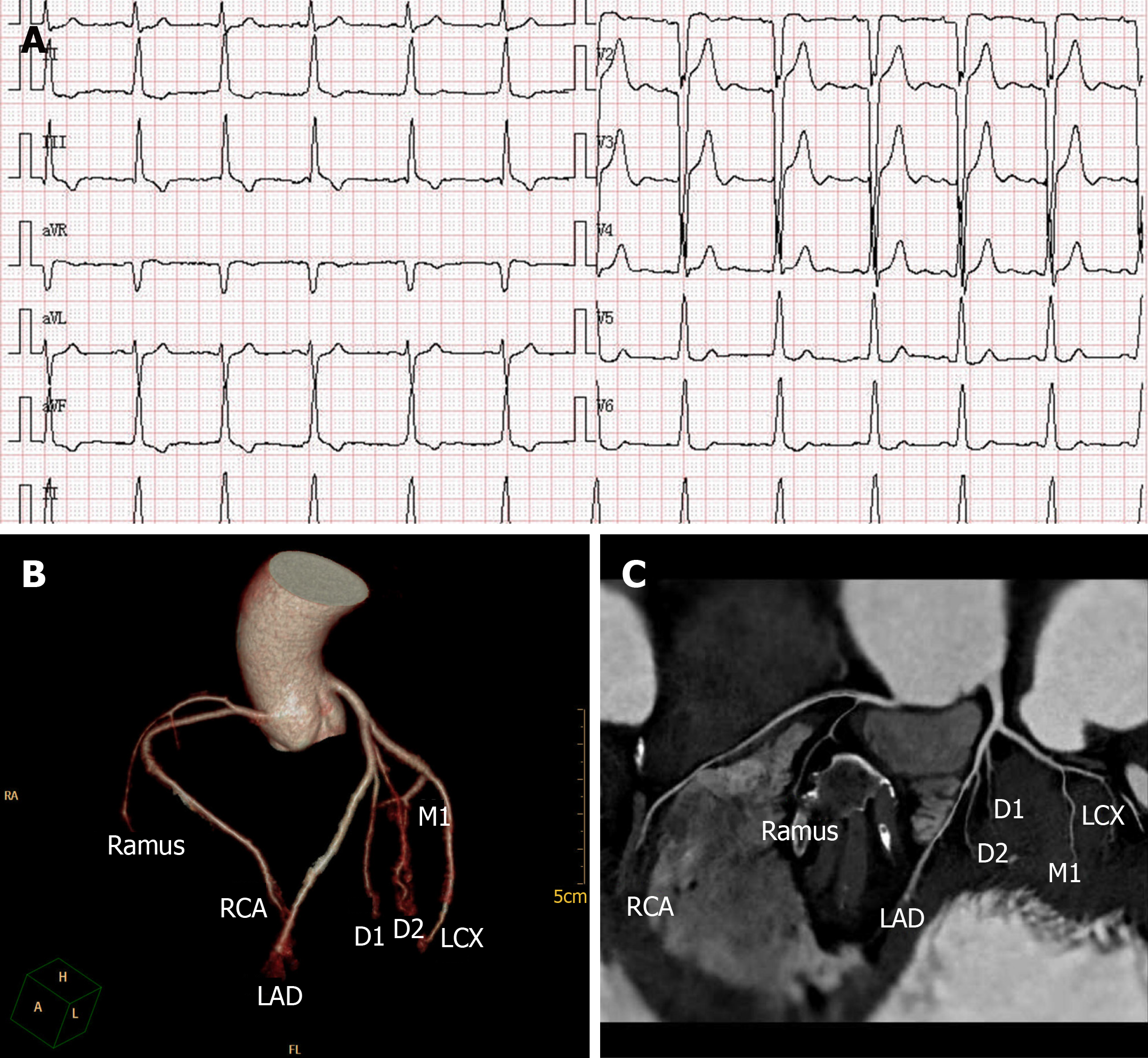
Figure 1 Electrocardiogram and coronary computed tomography angiography.
A: Twelve-lead electrocardiogram revealed a sinus rhythm with QS waves, an intraventricular conduction block (QRS duration, 118 ms), and a change of the ST-T wave in 2017. B and C: Coronary computed tomography angiography showed no abnormality in our patient. LAD: Left anterior descending; LCX: Left circumflex coronary artery; RCA: Right coronary artery; D1, D2: Diagonal branches; M1: Marginal branches.
- Citation: Chen YY, Yan H, Zhu JH. Successful treatment of systemic sclerosis complicated by ventricular tachycardia with a cardiac resynchronization therapy-defibrillator: A case report. World J Clin Cases 2020; 8(6): 1129-1136
- URL: https://www.wjgnet.com/2307-8960/full/v8/i6/1129.htm
- DOI: https://dx.doi.org/10.12998/wjcc.v8.i6.1129


