Copyright
©The Author(s) 2020.
World J Clin Cases. Mar 26, 2020; 8(6): 1116-1128
Published online Mar 26, 2020. doi: 10.12998/wjcc.v8.i6.1116
Published online Mar 26, 2020. doi: 10.12998/wjcc.v8.i6.1116
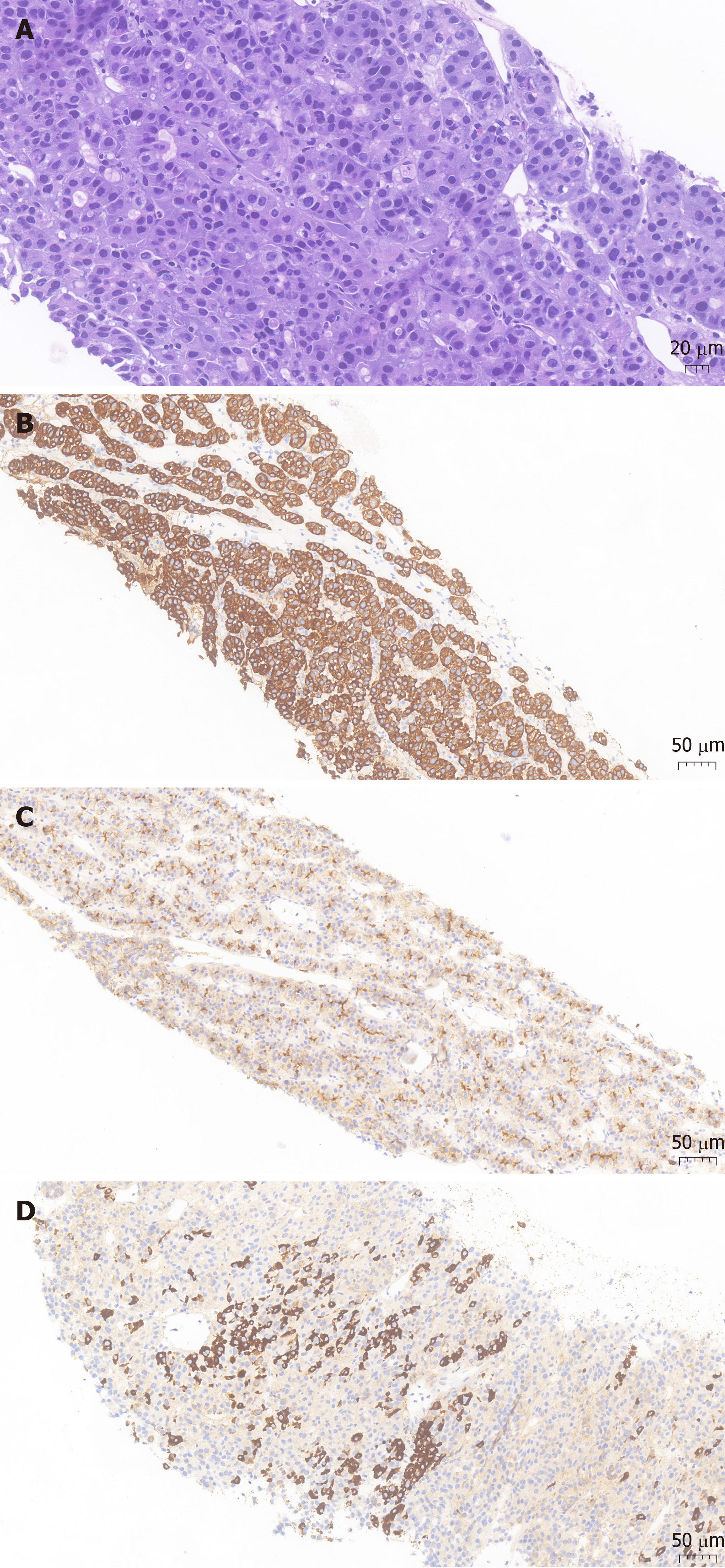
Figure 2 Pathological presentation of the patient.
A: Hematoxylin-eosin staining revealed heteromorphic neoplastic cells arranged in glandular, nested or striped patterns (magnification: × 200; scale bar: 20 μm); B: Immunohistochemistry showed tumor cell positivity for CK19 (magnification: × 100; scale bar: 50 μm); C: Immunohistochemistry showed tumor cell positivity for polyclonal carcinoembryonic antigen (magnification: × 100; scale bar: 50 μm); D: Immunohistochemistry showed tumor cell positivity for HepPar1 (magnification: × 200; scale bar: 50 μm).
- Citation: Zeng SX, Tan SW, Fong CJTH, Liang Q, Zhao BL, Liu K, Guo JX, Tao J. Hepatoid carcinoma of the pancreas: A case report and review of the literature. World J Clin Cases 2020; 8(6): 1116-1128
- URL: https://www.wjgnet.com/2307-8960/full/v8/i6/1116.htm
- DOI: https://dx.doi.org/10.12998/wjcc.v8.i6.1116


