Copyright
©The Author(s) 2020.
World J Clin Cases. Mar 26, 2020; 8(6): 1104-1107
Published online Mar 26, 2020. doi: 10.12998/wjcc.v8.i6.1104
Published online Mar 26, 2020. doi: 10.12998/wjcc.v8.i6.1104
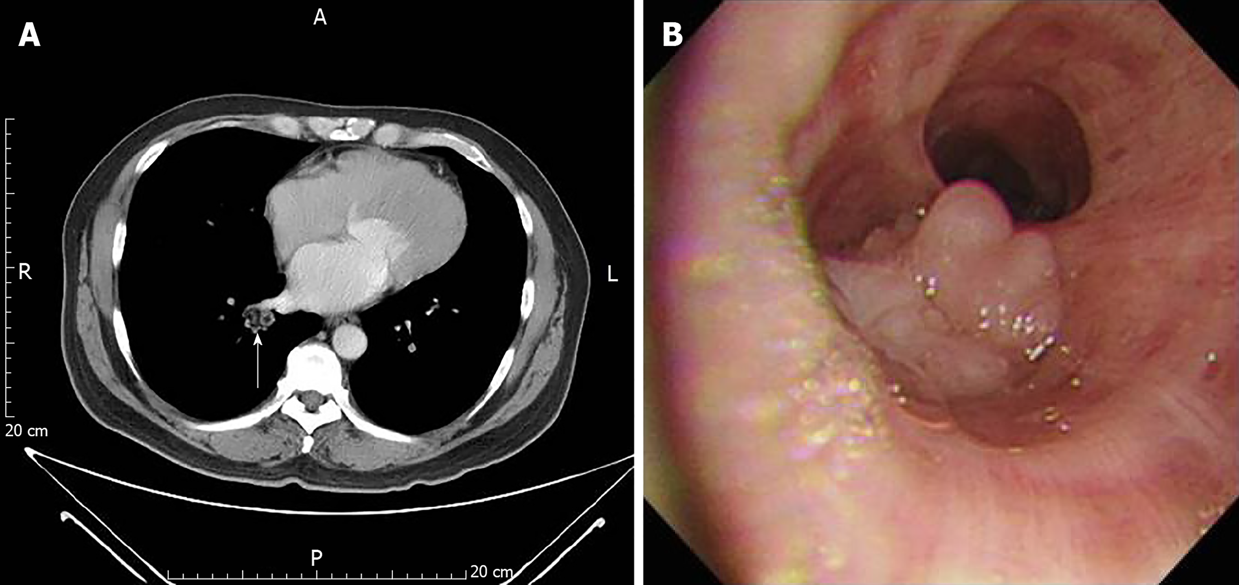
Figure 1 Computer tomography and bronchoscopy.
A: Contrast-enhanced computed tomography of the chest illustrates a nodule with lower density material over the right hilar region (arrow); B: Bronchoscopy showing an endobronchial lobulated protruded mass lesion in front of bronchi 9 and 10.
- Citation: Wu CW, Chen A, Huang TW. Diagnosis and management of glandular papilloma of lung: A case report. World J Clin Cases 2020; 8(6): 1104-1107
- URL: https://www.wjgnet.com/2307-8960/full/v8/i6/1104.htm
- DOI: https://dx.doi.org/10.12998/wjcc.v8.i6.1104


