Copyright
©The Author(s) 2020.
World J Clin Cases. Mar 26, 2020; 8(6): 1056-1064
Published online Mar 26, 2020. doi: 10.12998/wjcc.v8.i6.1056
Published online Mar 26, 2020. doi: 10.12998/wjcc.v8.i6.1056
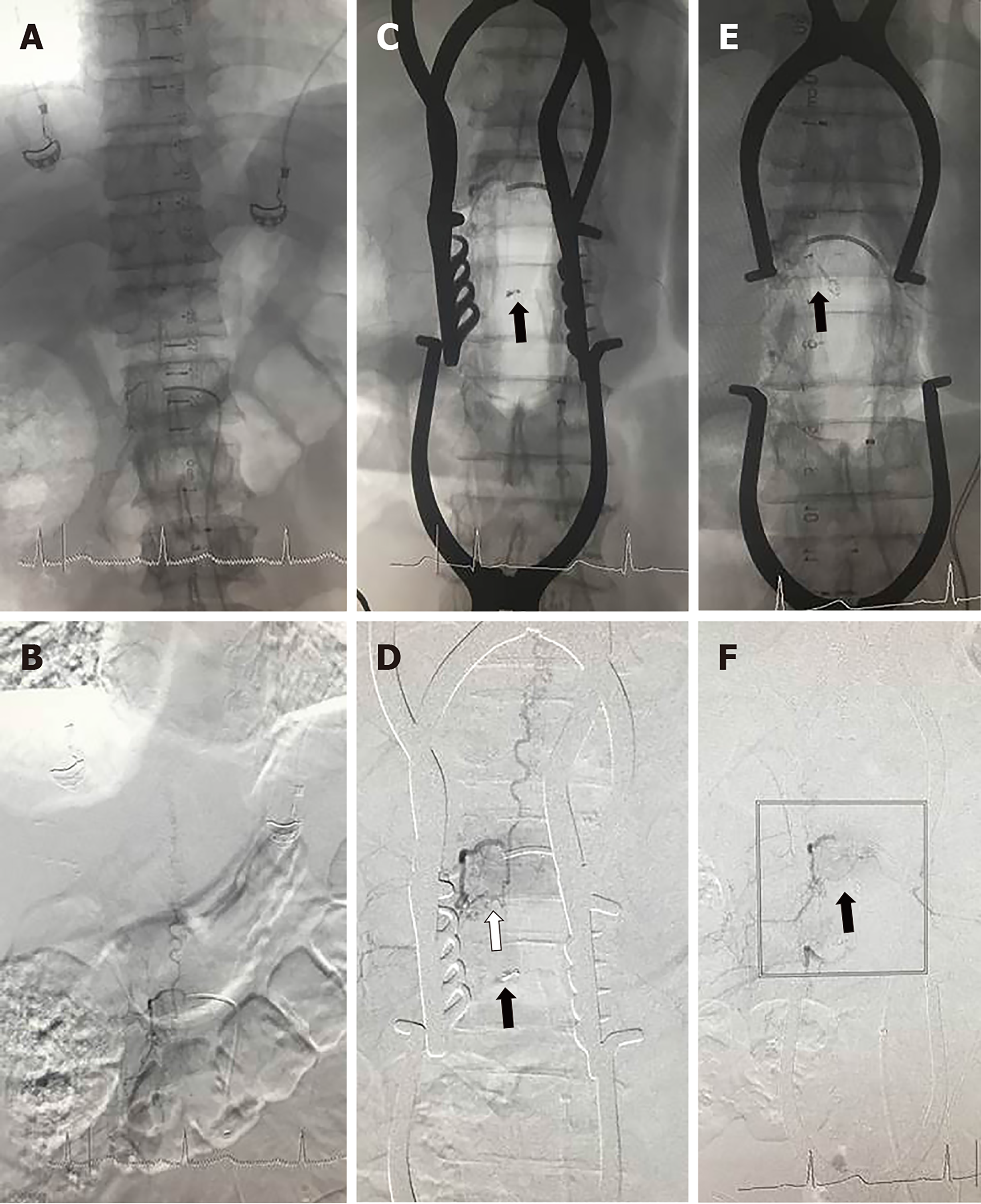
Figure 2 Intraoperative angiograms obtained in Case 11 with an L-1 lumbar SDAVF treated in hybrid OR.
A and B: Preoperative MASK and digital subtraction angiography (DSA) images displayed that the location of the spinal dural arteriovenous fistula was predicted to be in the dura at the right L-1 dorsal root; C and D: During the operation, a suspected feeding artery was found initially and clipped temporarily under the microscope; however, the first intraoperative MASK and DSA images displayed that the fistula (white arrow) was still visualized and that the microvascular clip (black arrow) was below the right fistula; E and F: The second intraoperative MASK and DSA images showed that the spinal dural arteriovenous fistula had completely disappeared, and the microvascular clip (black arrow) was adjusted at the right fistula. DSA: Digital subtraction angiography.
- Citation: Zhang N, Xin WQ. Application of hybrid operating rooms for treating spinal dural arteriovenous fistula. World J Clin Cases 2020; 8(6): 1056-1064
- URL: https://www.wjgnet.com/2307-8960/full/v8/i6/1056.htm
- DOI: https://dx.doi.org/10.12998/wjcc.v8.i6.1056


