Copyright
©The Author(s) 2020.
World J Clin Cases. Mar 6, 2020; 8(5): 963-970
Published online Mar 6, 2020. doi: 10.12998/wjcc.v8.i5.963
Published online Mar 6, 2020. doi: 10.12998/wjcc.v8.i5.963
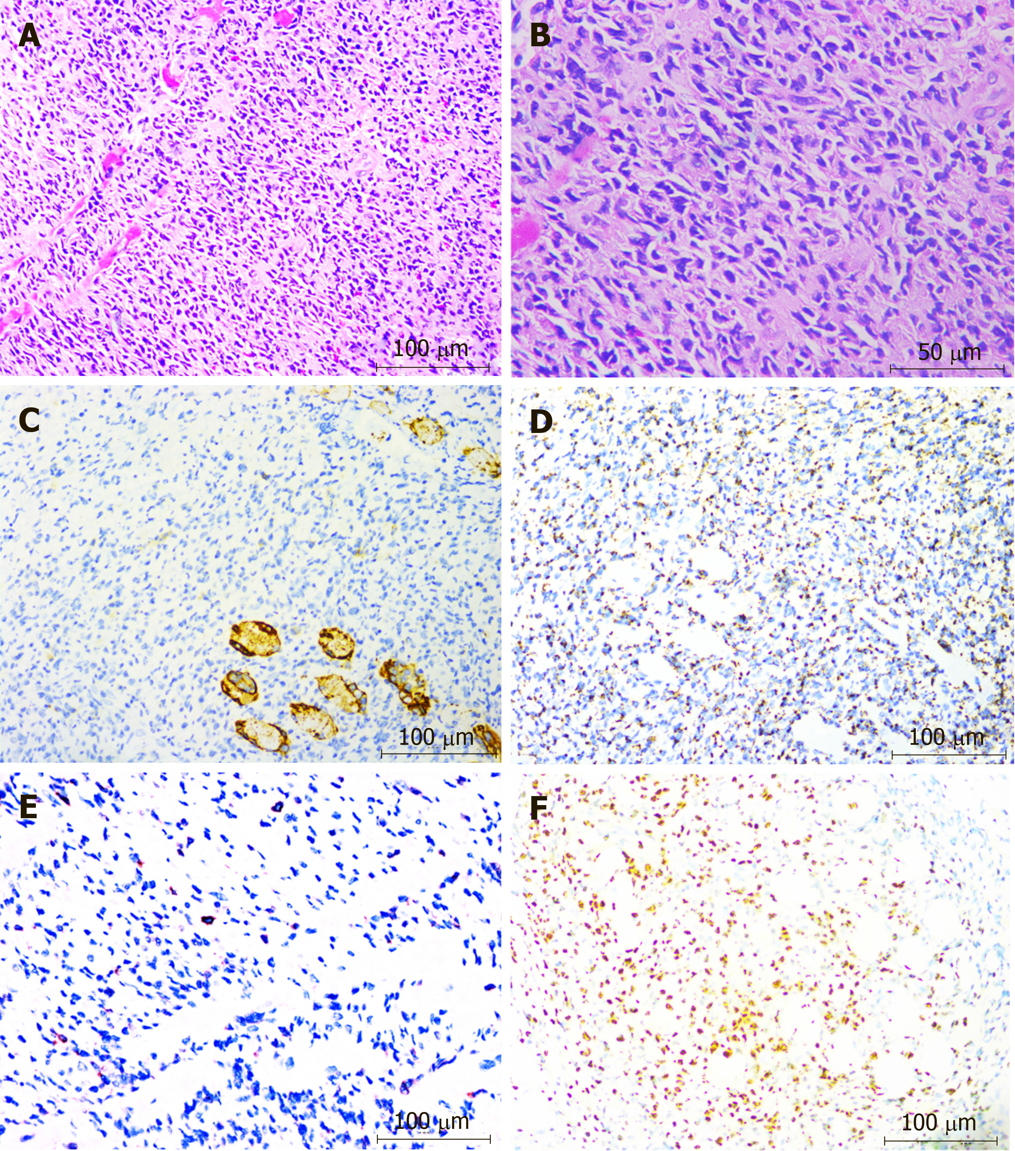
Figure 2 Immunohistochemical staining.
A and B: Histological findings of the pharyngeal wall (hematoxylin-eosin: panel A, × 200; panel B, × 400), showing diffuse infiltration with atypical lymphoid cells; C: Immunohistochemical staining for CD56 was negative (× 200); D-F: Immunohistochemical staining was positive for TIA-1 (panel D, ×200), CD30 (panel E, × 200) and EBER (panel F, × 200).
- Citation: Liu LH, Huang Q, Liu YH, Yang J, Fu H, Jin L. Muscular involvement of extranodal natural killer/T cell lymphoma misdiagnosed as polymyositis: A case report and review of literature. World J Clin Cases 2020; 8(5): 963-970
- URL: https://www.wjgnet.com/2307-8960/full/v8/i5/963.htm
- DOI: https://dx.doi.org/10.12998/wjcc.v8.i5.963


