Copyright
©The Author(s) 2020.
World J Clin Cases. Mar 6, 2020; 8(5): 939-945
Published online Mar 6, 2020. doi: 10.12998/wjcc.v8.i5.939
Published online Mar 6, 2020. doi: 10.12998/wjcc.v8.i5.939
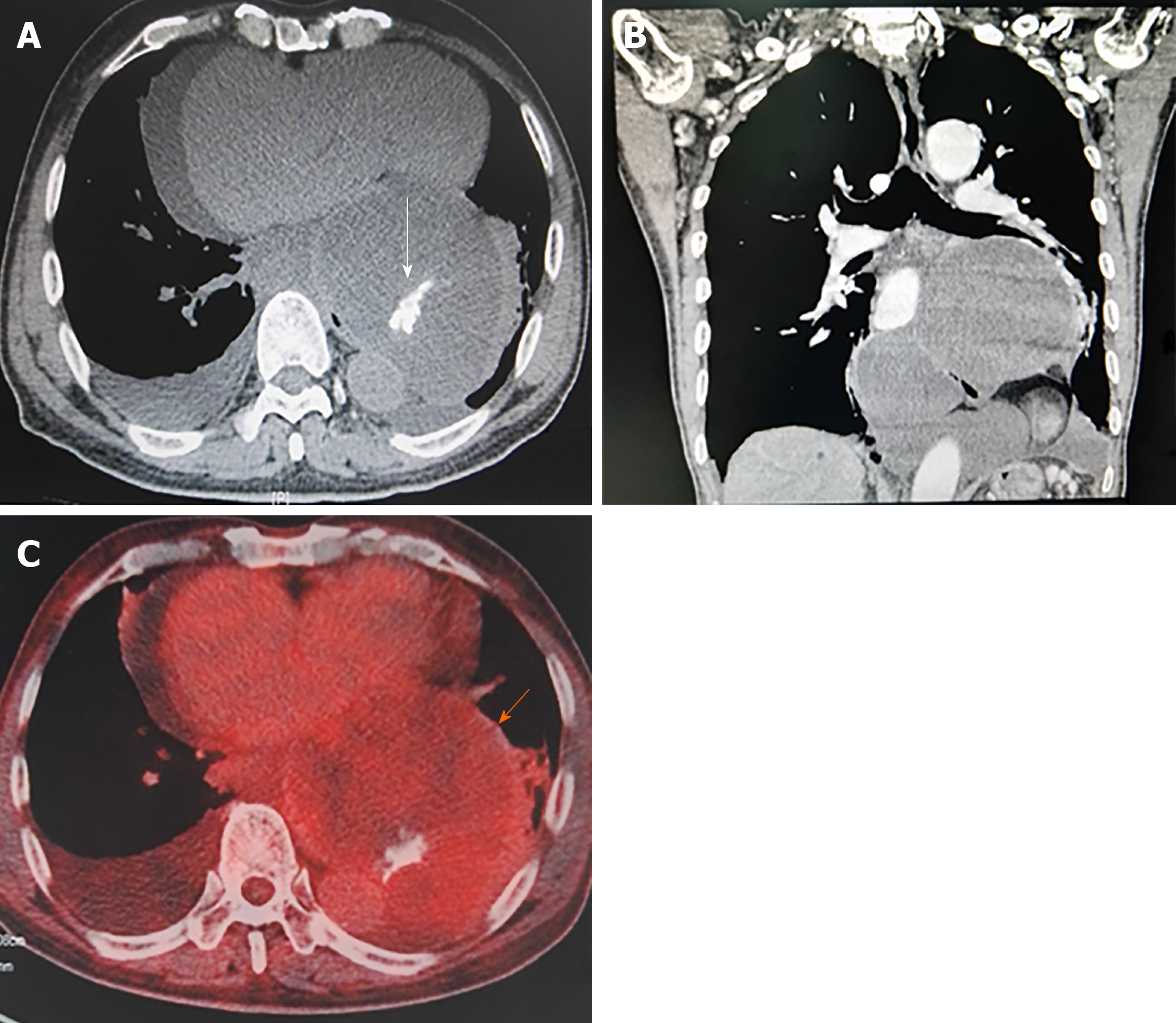
Figure 1 Imaging findings.
A and B: Chest contrast-enhanced computed tomography scans showed several cystic-solid encapsulated masses in posterior-inferior mediastinum with well-defined margins, fatty density, soft tissue density, calcification and slight enhancement; C: 18F-fluorodeoxyglucose PET showed the maximum standardized uptake value (SUVave) of 1.8.
- Citation: Chen HG, Zhang K, Wu WB, Wu YH, Zhang J, Gu LJ, Li XJ. Combining surgery with 125I brachytherapy for recurrent mediastinal dedifferentiated liposarcoma: A case report and review of literature. World J Clin Cases 2020; 8(5): 939-945
- URL: https://www.wjgnet.com/2307-8960/full/v8/i5/939.htm
- DOI: https://dx.doi.org/10.12998/wjcc.v8.i5.939


