Copyright
©The Author(s) 2020.
World J Clin Cases. Mar 6, 2020; 8(5): 912-921
Published online Mar 6, 2020. doi: 10.12998/wjcc.v8.i5.912
Published online Mar 6, 2020. doi: 10.12998/wjcc.v8.i5.912
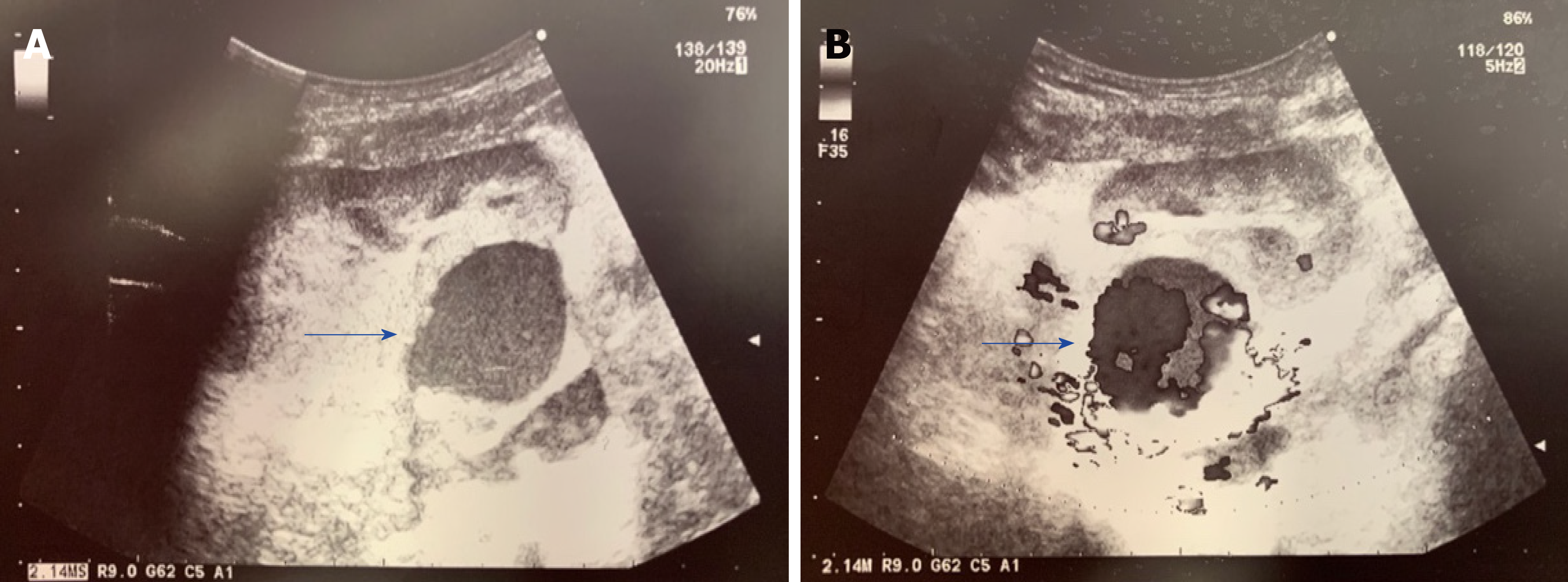
Figure 1 Color Doppler ultrasound scan of the transplanted kidney.
A: Rounded, 3.8 cm-sized, hypoechoic mass localized between the hilum of the allograft and the iliac vessels (blue arrow); B: Turbulent intra-lesional flow (yin-yang sign) along the renal allograft artery (blue arrow).
- Citation: Bindi M, Ferraresso M, De Simeis ML, Raison N, Clementoni L, Delbue S, Perego M, Favi E. Allograft artery mycotic aneurysm after kidney transplantation: A case report and review of literature. World J Clin Cases 2020; 8(5): 912-921
- URL: https://www.wjgnet.com/2307-8960/full/v8/i5/912.htm
- DOI: https://dx.doi.org/10.12998/wjcc.v8.i5.912


