Copyright
©The Author(s) 2020.
World J Clin Cases. Feb 26, 2020; 8(4): 838-847
Published online Feb 26, 2020. doi: 10.12998/wjcc.v8.i4.838
Published online Feb 26, 2020. doi: 10.12998/wjcc.v8.i4.838
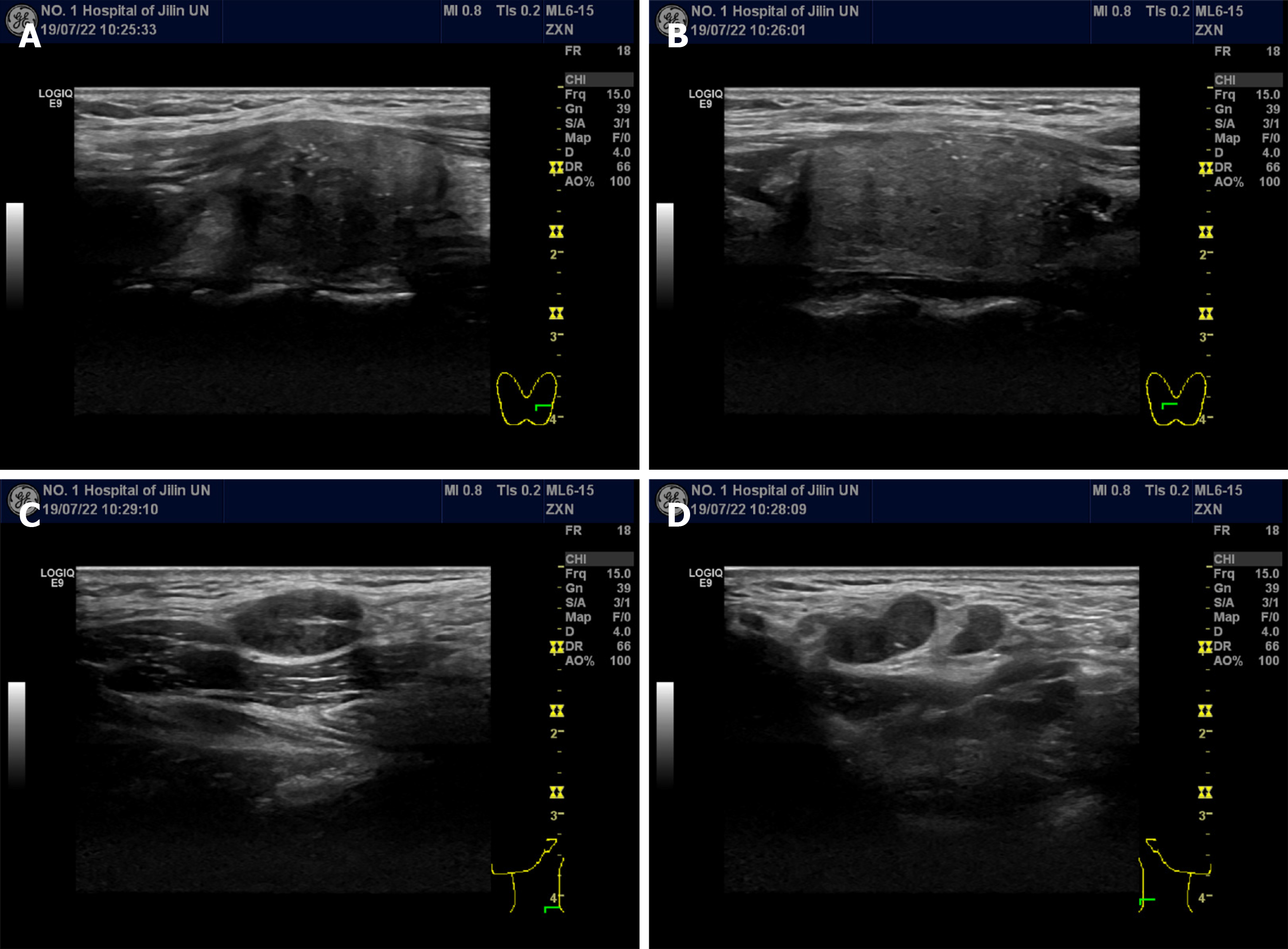
Figure 1 Ultrasonography of the thyroid and lateral neck.
A and B: Left (A) and right (B) thyroid lobes; solid, hypoechoic thyroid nodules with irregular margins could be observed; C and D: Left (C) and right (D) lateral neck; enlarged metastatic lateral lymph nodes could be observed.
- Citation: Zhang YY, Xue S, Wang ZM, Jin MS, Chen ZP, Chen G, Zhang Q. Thyroid metastasis from breast cancer presenting with enlarged lateral cervical lymph nodes: A case report. World J Clin Cases 2020; 8(4): 838-847
- URL: https://www.wjgnet.com/2307-8960/full/v8/i4/838.htm
- DOI: https://dx.doi.org/10.12998/wjcc.v8.i4.838


