Copyright
©The Author(s) 2020.
World J Clin Cases. Feb 26, 2020; 8(4): 806-814
Published online Feb 26, 2020. doi: 10.12998/wjcc.v8.i4.806
Published online Feb 26, 2020. doi: 10.12998/wjcc.v8.i4.806
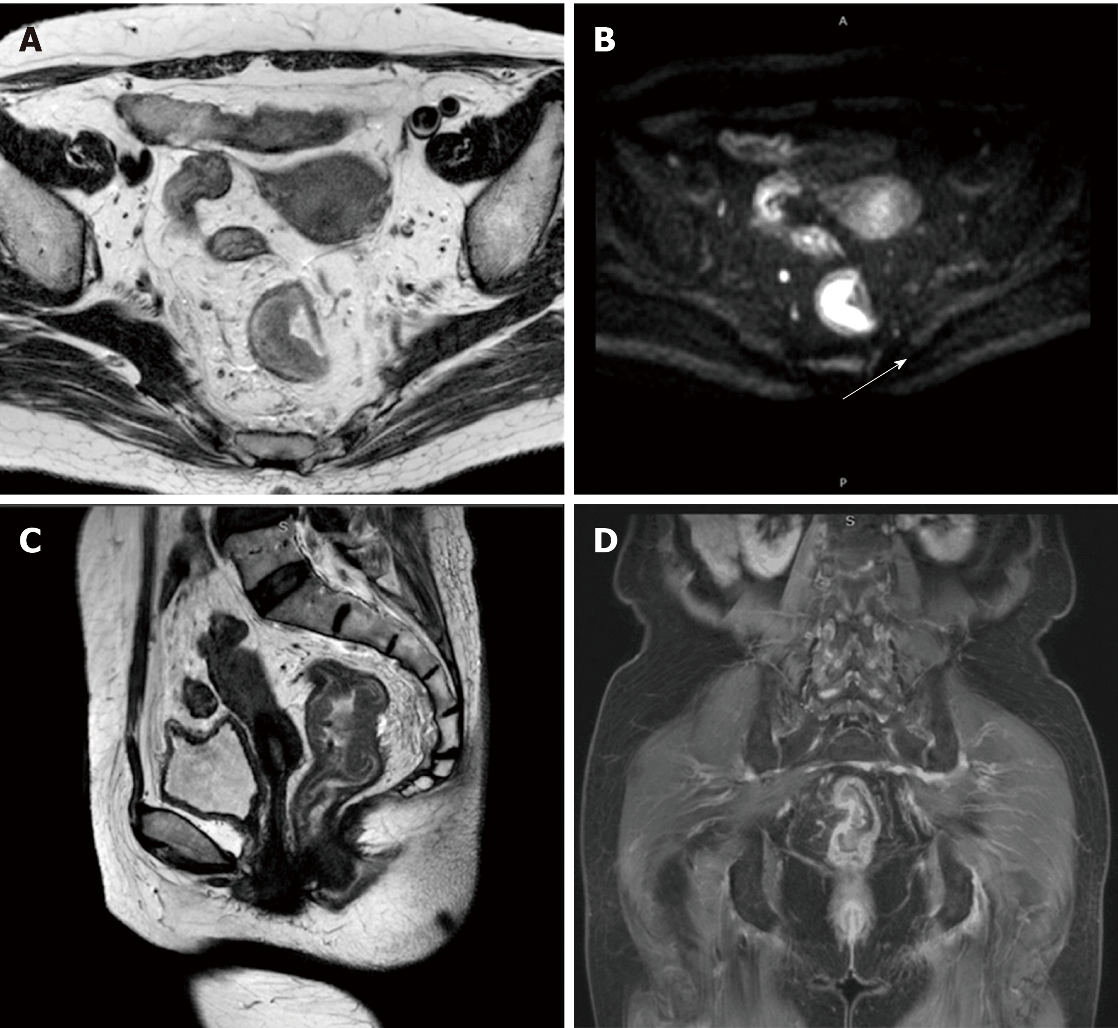
Figure 5 Pelvic magnetic resonance imaging.
A: T2WI image revealing that a soft tissue mass was irregularly thickened and was seen to protrude into the lumen; B: Axial DWI image showing a high signal of the mass (white arrow); C: Pelvic magnetic resonance imaging showing rectal wall thickening (coronal view); D: Pelvic magnetic resonance imaging showing rectal wall thickening. (sagittal view).
- Citation: Chen T, Que YT, Zhang YH, Long FY, Li Y, Huang X, Wang YN, Hu YF, Yu J, Li GX. Using Materialise's interactive medical image control system to reconstruct a model of a patient with rectal cancer and situs inversus totalis: A case report. World J Clin Cases 2020; 8(4): 806-814
- URL: https://www.wjgnet.com/2307-8960/full/v8/i4/806.htm
- DOI: https://dx.doi.org/10.12998/wjcc.v8.i4.806


