Copyright
©The Author(s) 2020.
World J Clin Cases. Feb 26, 2020; 8(4): 806-814
Published online Feb 26, 2020. doi: 10.12998/wjcc.v8.i4.806
Published online Feb 26, 2020. doi: 10.12998/wjcc.v8.i4.806
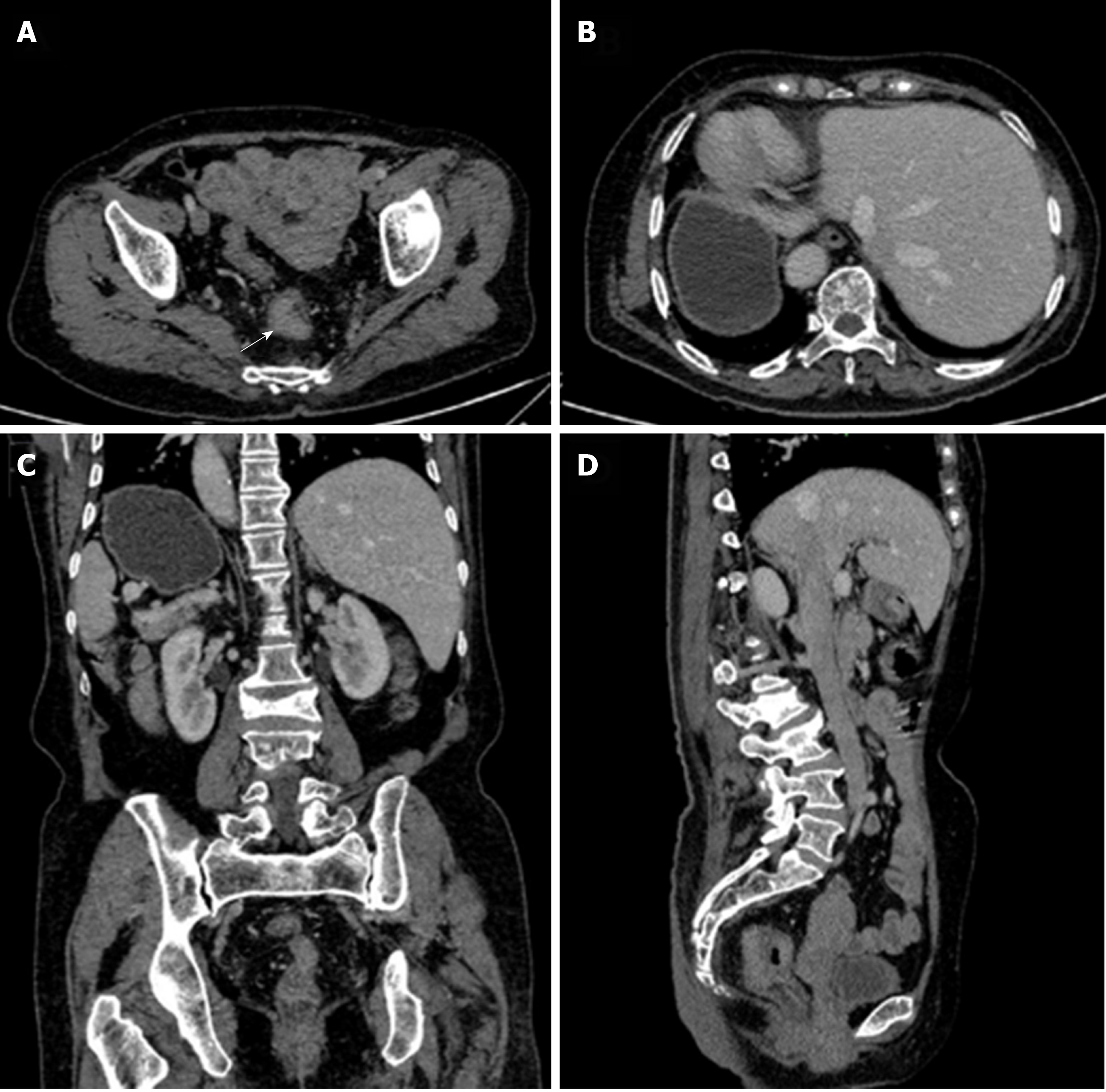
Figure 4 Abdominopelvic computed tomography images.
A: Computed tomography (CT) image revealing that a soft tissue mass was irregularly infiltrated into the serous membrane (white arrow). The mass was seen to protrude into the lumen, with metastatic lymph nodes detected around; B: CT image further revealing the complete transposition of the abdominal viscera; C: CT image disclosing complete transposition of the abdominal viscera (coronal view); D: CT image disclosing complete transposition of the abdominal viscera (sagittal view).
- Citation: Chen T, Que YT, Zhang YH, Long FY, Li Y, Huang X, Wang YN, Hu YF, Yu J, Li GX. Using Materialise's interactive medical image control system to reconstruct a model of a patient with rectal cancer and situs inversus totalis: A case report. World J Clin Cases 2020; 8(4): 806-814
- URL: https://www.wjgnet.com/2307-8960/full/v8/i4/806.htm
- DOI: https://dx.doi.org/10.12998/wjcc.v8.i4.806


