Copyright
©The Author(s) 2020.
World J Clin Cases. Feb 6, 2020; 8(3): 630-637
Published online Feb 6, 2020. doi: 10.12998/wjcc.v8.i3.630
Published online Feb 6, 2020. doi: 10.12998/wjcc.v8.i3.630
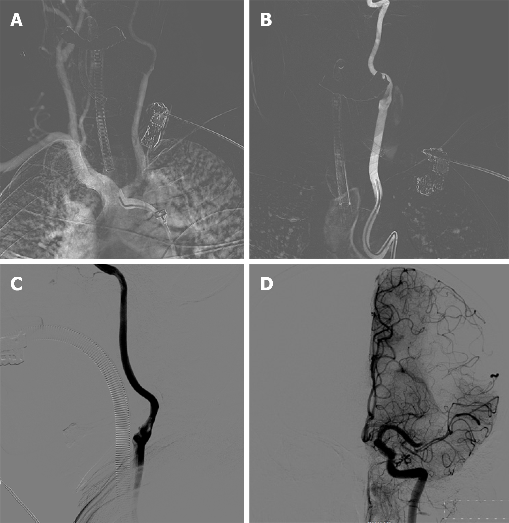
Figure 2 Intracranial angiography findings.
A: Subaortic arch angiography shows a type III aortic arch where the left common carotid artery shares the main trunk with the brachiocephalic trunk; B: Simmon 2 catheterangiography showing that the left external carotid artery was not visible, and the left internal carotid artery showing slow blood flow; C: There was a thrombus at the beginning of the left internal carotid artery; D: The upper trunk of the left middle cerebral artery was occluded, but the anterior cerebral artery compensated for the blood supply of the middle cerebral artery via the lateral branches of the meninges.
- Citation: Zhang M, Hao JH, Lin K, Cui QK, Zhang LY. Combined surgical and interventional treatment of tandem carotid artery and middle cerebral artery embolus: A case report. World J Clin Cases 2020; 8(3): 630-637
- URL: https://www.wjgnet.com/2307-8960/full/v8/i3/630.htm
- DOI: https://dx.doi.org/10.12998/wjcc.v8.i3.630


