Copyright
©The Author(s) 2020.
World J Clin Cases. Feb 6, 2020; 8(3): 568-576
Published online Feb 6, 2020. doi: 10.12998/wjcc.v8.i3.568
Published online Feb 6, 2020. doi: 10.12998/wjcc.v8.i3.568
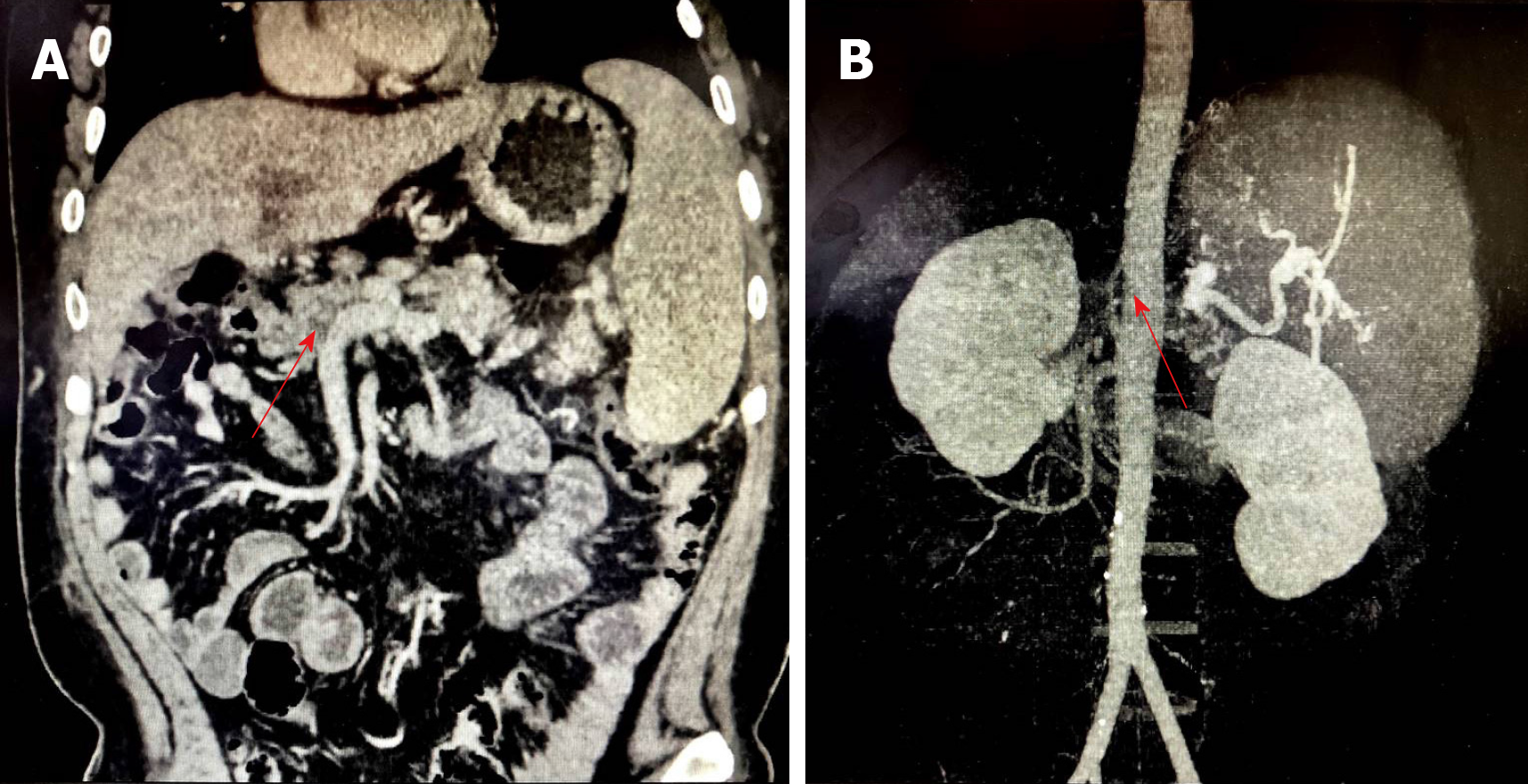
Figure 1 Preoperative abdominal imaging of the patient.
A: From the 5th year after primary liver transplantation, abdominal enhanced computed tomography showed portal vein thrombosis with cavernous degeneration of the portal vein (red arrow); B: Abdominal enhanced computed tomography showed portal vein thrombosis with occlusion of the hepatic artery (red arrow).
- Citation: Li J, Guo QJ, Jiang WT, Zheng H, Shen ZY. Complex liver retransplantation to treat graft loss due to long-term biliary tract complication after liver transplantation: A case report. World J Clin Cases 2020; 8(3): 568-576
- URL: https://www.wjgnet.com/2307-8960/full/v8/i3/568.htm
- DOI: https://dx.doi.org/10.12998/wjcc.v8.i3.568


