Copyright
©The Author(s) 2020.
World J Clin Cases. Feb 6, 2020; 8(3): 552-559
Published online Feb 6, 2020. doi: 10.12998/wjcc.v8.i3.552
Published online Feb 6, 2020. doi: 10.12998/wjcc.v8.i3.552
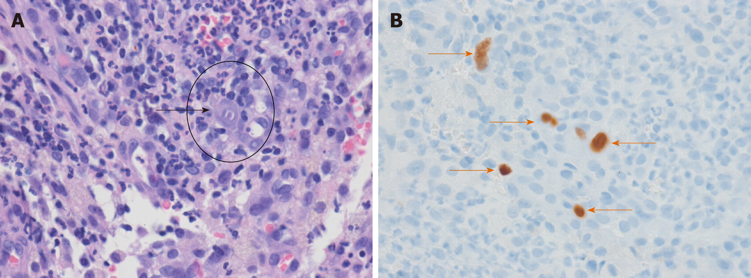
Figure 3 Pathology findings of hematoxylin-eosin and immunohistochemical stained biopsy sections.
A: The black arrow shows cytomegalovirus inclusion bodies (HE staining, × 400); B: Orange arrows show cytomegalovirus-positive cells (immunohistochemical staining, × 400).
- Citation: Cho JH, Choi JH. Cytomegalovirus ileo-pancolitis presenting as toxic megacolon in an immunocompetent patient: A case report. World J Clin Cases 2020; 8(3): 552-559
- URL: https://www.wjgnet.com/2307-8960/full/v8/i3/552.htm
- DOI: https://dx.doi.org/10.12998/wjcc.v8.i3.552


