Copyright
©The Author(s) 2020.
World J Clin Cases. Feb 6, 2020; 8(3): 522-526
Published online Feb 6, 2020. doi: 10.12998/wjcc.v8.i3.522
Published online Feb 6, 2020. doi: 10.12998/wjcc.v8.i3.522
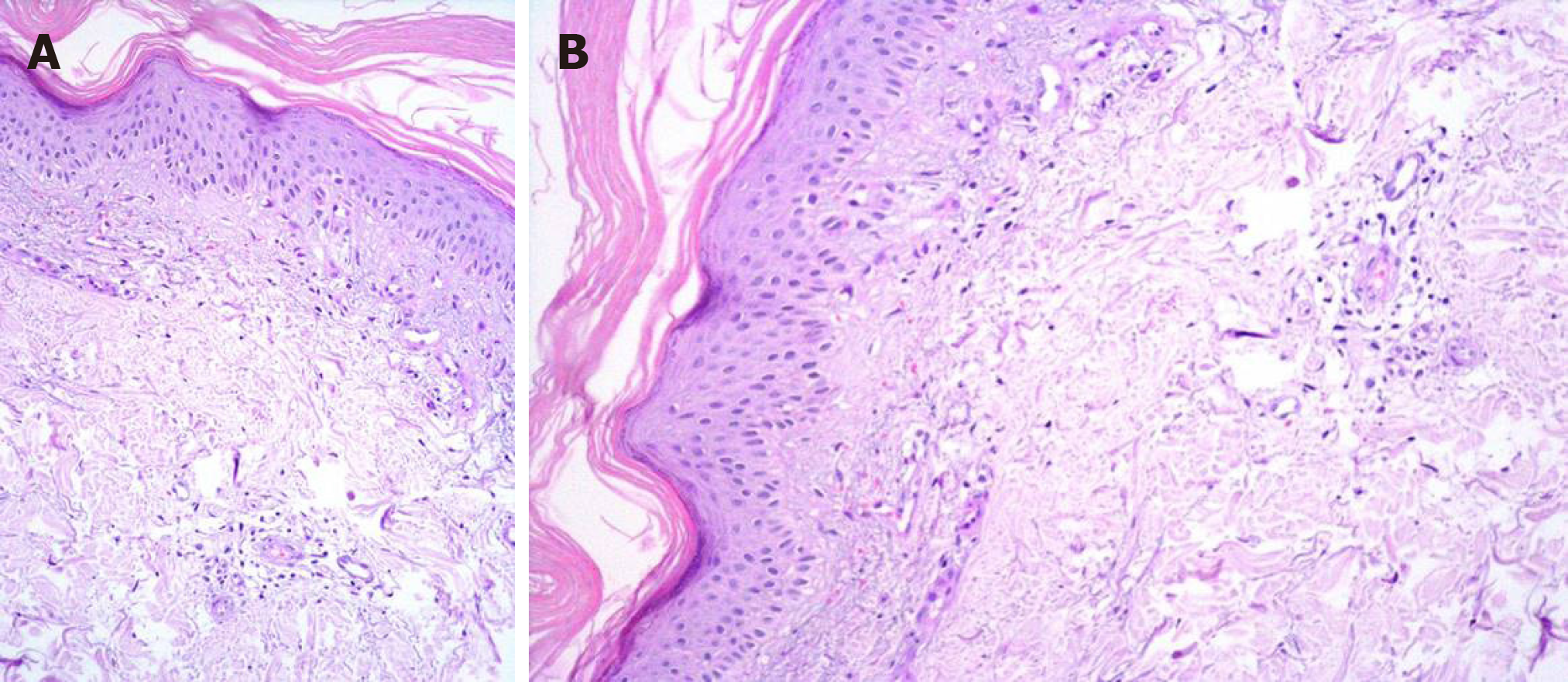
Figure 3 A dermal biopsy was performed and orthokeratotic epidermis with lymphocytic infiltration was observed in the pathologic specimen.
A: Perivascular lymphocytes (Hematoxylin-eosin staining, × 20); B: Perivascular lymphocytes (Hematoxylin-eosin staining, × 20).
- Citation: Yilmaz M, Celik U, Hascicek S. Radiation recall dermatitis with dabrafenib and trametinib: A case report. World J Clin Cases 2020; 8(3): 522-526
- URL: https://www.wjgnet.com/2307-8960/full/v8/i3/522.htm
- DOI: https://dx.doi.org/10.12998/wjcc.v8.i3.522


