Copyright
©The Author(s) 2020.
World J Clin Cases. Feb 6, 2020; 8(3): 504-516
Published online Feb 6, 2020. doi: 10.12998/wjcc.v8.i3.504
Published online Feb 6, 2020. doi: 10.12998/wjcc.v8.i3.504
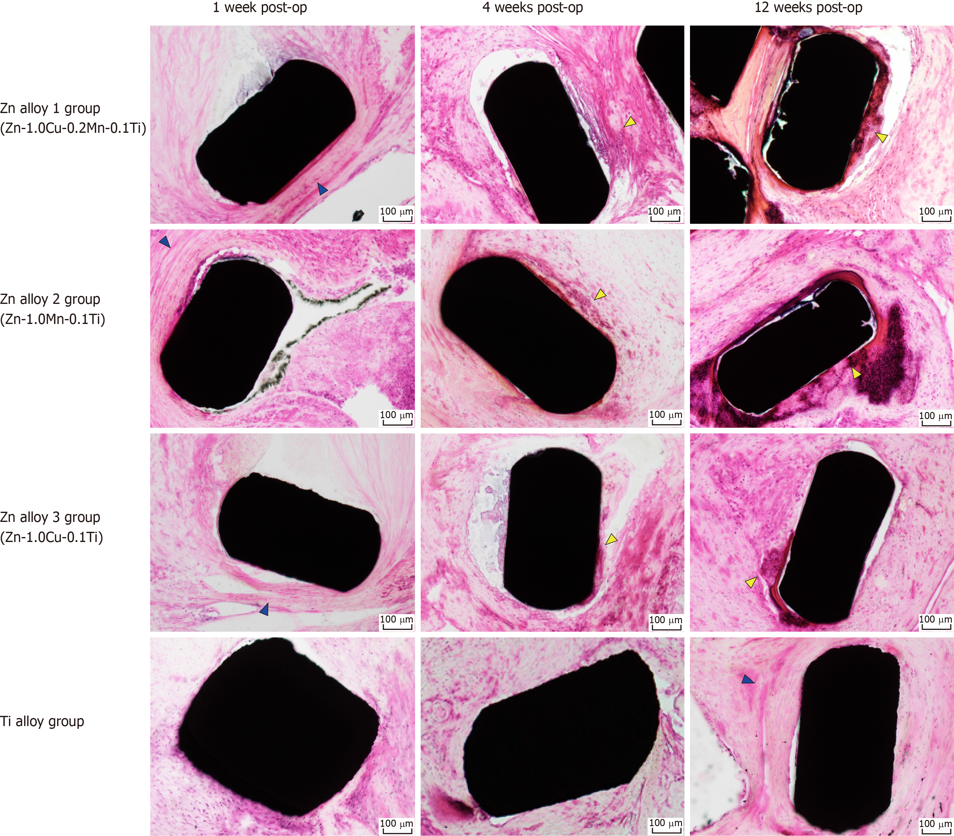
Figure 7 Histopathological images of rabbit gastric tissues surrounding Zn alloy and Ti alloy staples after hematoxylin–eosin staining.
Blue arrows indicate fibrous layers of tissue (observed around both, the Ti alloy and Zn alloy staples), and yellow arrows indicate inflammatory cell infiltrations (localized around Zn alloy staples only).
- Citation: Amano H, Miyake K, Hinoki A, Yokota K, Kinoshita F, Nakazawa A, Tanaka Y, Seto Y, Uchida H. Novel zinc alloys for biodegradable surgical staples. World J Clin Cases 2020; 8(3): 504-516
- URL: https://www.wjgnet.com/2307-8960/full/v8/i3/504.htm
- DOI: https://dx.doi.org/10.12998/wjcc.v8.i3.504


