Copyright
©The Author(s) 2020.
World J Clin Cases. Dec 26, 2020; 8(24): 6537-6545
Published online Dec 26, 2020. doi: 10.12998/wjcc.v8.i24.6537
Published online Dec 26, 2020. doi: 10.12998/wjcc.v8.i24.6537
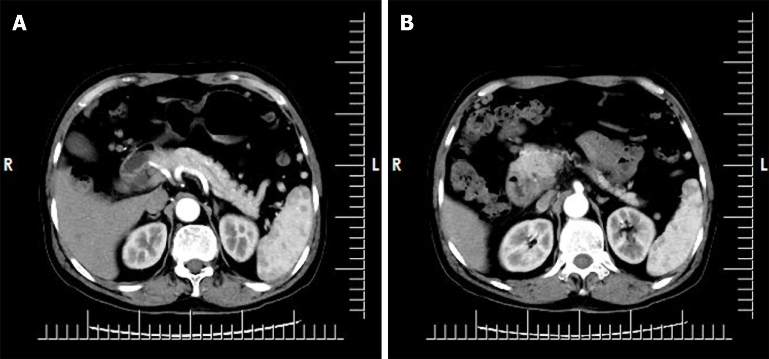
Figure 1 Upper abdominal enhanced computed tomography at Beijing Dong Fang Hospital.
A: The wall of the descending part of the duodenum showed local thickening, with an iso-low density shadow and blurred edge; B: Enhanced scan showed obvious non-uniform enhancement. The impression diagnosis was space-occupying lesion in the descending part of the duodenum, and malignancy was considered.
- Citation: Zhang Y, Shi XJ, Zhang XC, Zhao XJ, Li JX, Wang LH, Xie CE, Liu YY, Wang YL. Primary duodenal tuberculosis misdiagnosed as tumor by imaging examination: A case report. World J Clin Cases 2020; 8(24): 6537-6545
- URL: https://www.wjgnet.com/2307-8960/full/v8/i24/6537.htm
- DOI: https://dx.doi.org/10.12998/wjcc.v8.i24.6537


