Copyright
©The Author(s) 2020.
World J Clin Cases. Dec 26, 2020; 8(24): 6524-6528
Published online Dec 26, 2020. doi: 10.12998/wjcc.v8.i24.6524
Published online Dec 26, 2020. doi: 10.12998/wjcc.v8.i24.6524
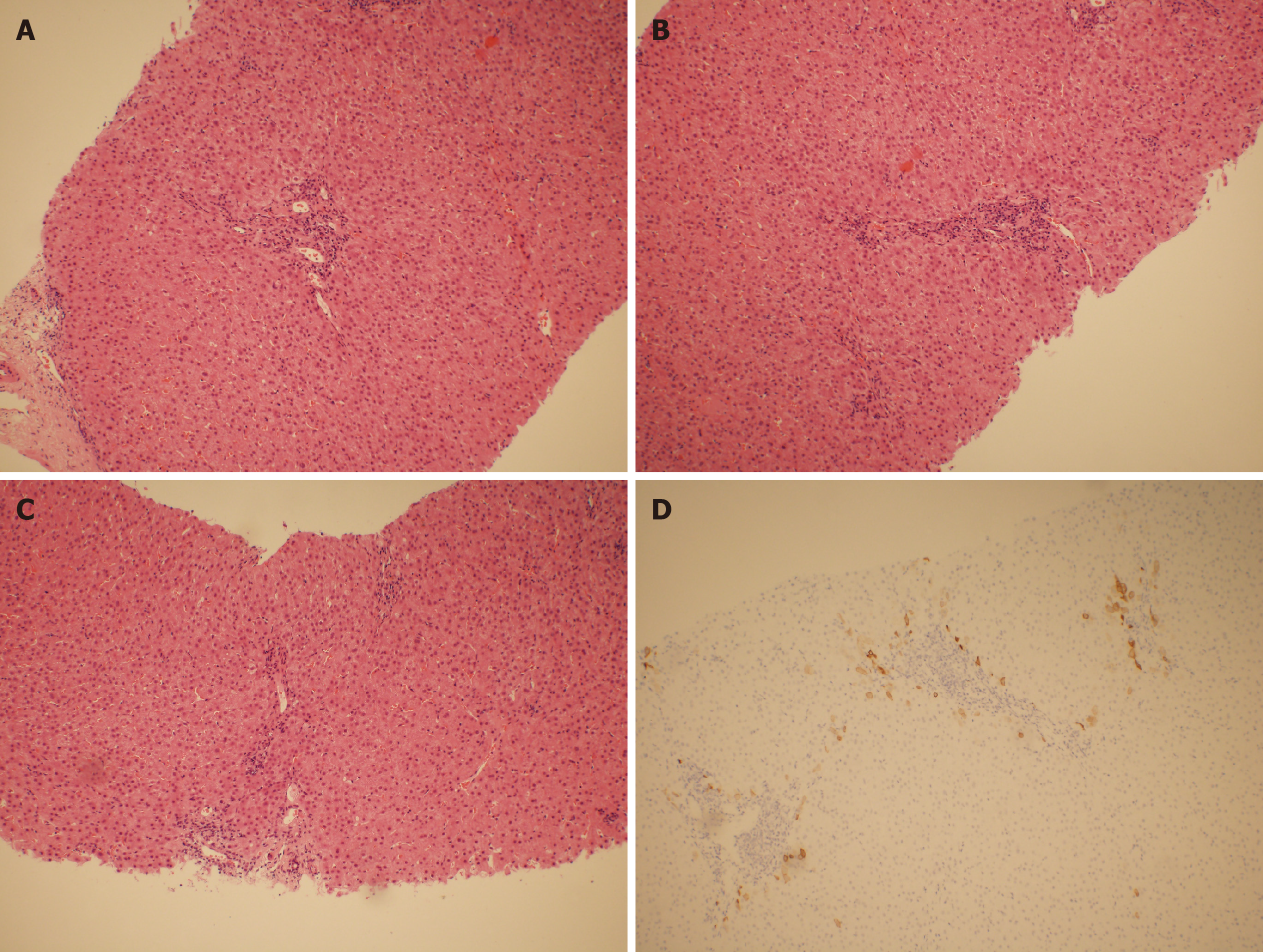
Figure 1 Pathology and immunohistochemical staining.
A-C: Liver pathology staining showing plasma cell infiltration and focal interfacial inflammation in the confluence area and mild interfacial inflammation; D: Immunohistochemical staining of cytokeratin 7 showing that most of the intrahepatic bile ducts were missing (> 50%).
- Citation: Zhang XC, Wang D, Li X, Hu YL, Wang C. Idiopathic adulthood ductopenia with elevated transaminase only: A case report. World J Clin Cases 2020; 8(24): 6524-6528
- URL: https://www.wjgnet.com/2307-8960/full/v8/i24/6524.htm
- DOI: https://dx.doi.org/10.12998/wjcc.v8.i24.6524


