Copyright
©The Author(s) 2020.
World J Clin Cases. Dec 26, 2020; 8(24): 6487-6498
Published online Dec 26, 2020. doi: 10.12998/wjcc.v8.i24.6487
Published online Dec 26, 2020. doi: 10.12998/wjcc.v8.i24.6487
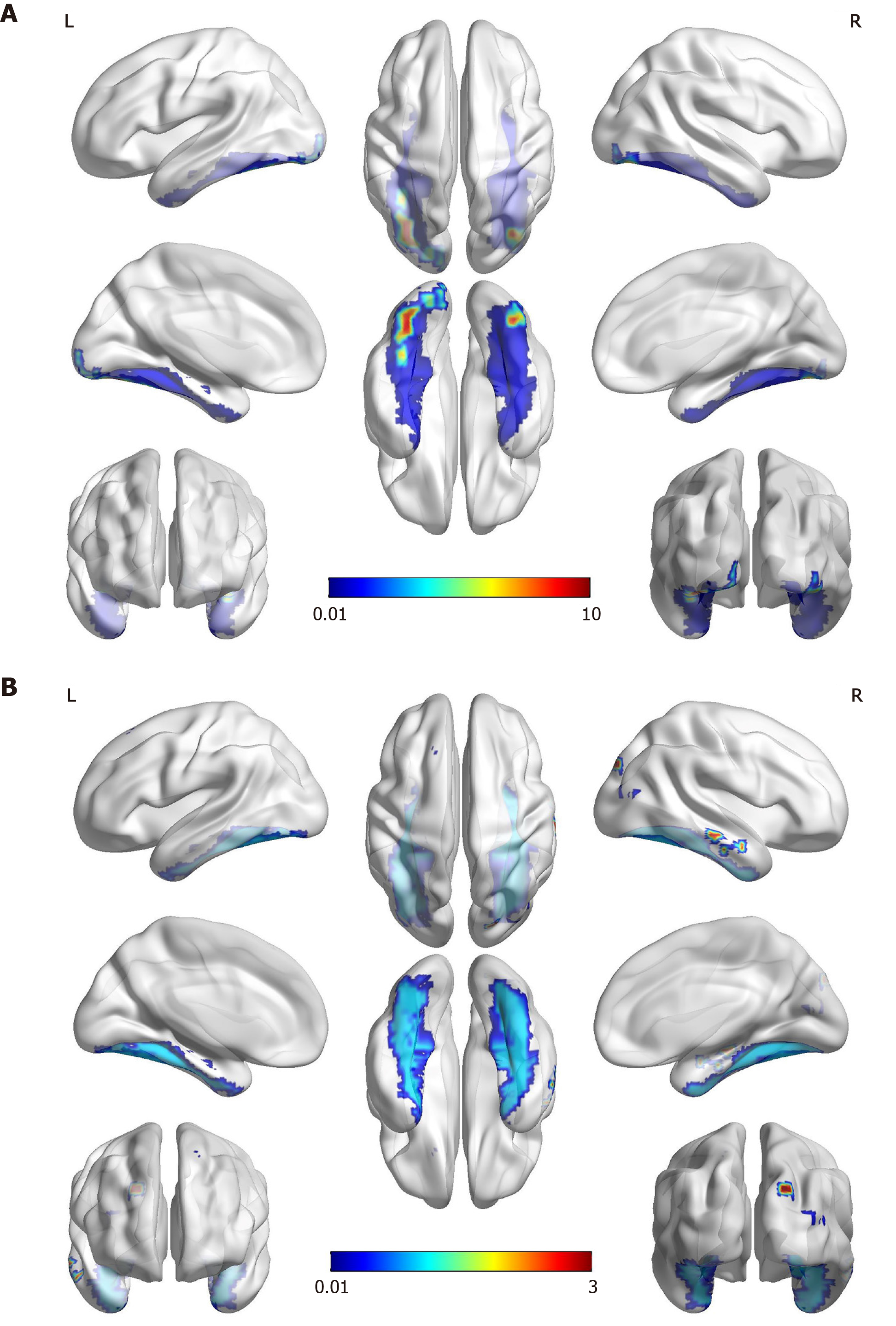
Figure 3 Functional magnetic resonance imaging.
A: The famous faces task performed two months after the onset of stroke showed activation in the anterior of the bilateral fusiform gyrus that was more significant on the left (red and yellow). The areas of activation are correlated with the fusiform gyrus (blue), which most likely corresponds to the “fusiform face area”; B: The object/landscape task demonstrated activation in the area of the right occipital lobe, which is distinct from the face recognition area.
- Citation: Yuan Y, Huang F, Gao ZH, Cai WC, Xiao JX, Yang YE, Zhu PL. Delayed diagnosis of prosopagnosia following a hemorrhagic stroke in an elderly man: A case report. World J Clin Cases 2020; 8(24): 6487-6498
- URL: https://www.wjgnet.com/2307-8960/full/v8/i24/6487.htm
- DOI: https://dx.doi.org/10.12998/wjcc.v8.i24.6487


