Copyright
©The Author(s) 2020.
World J Clin Cases. Dec 26, 2020; 8(24): 6487-6498
Published online Dec 26, 2020. doi: 10.12998/wjcc.v8.i24.6487
Published online Dec 26, 2020. doi: 10.12998/wjcc.v8.i24.6487
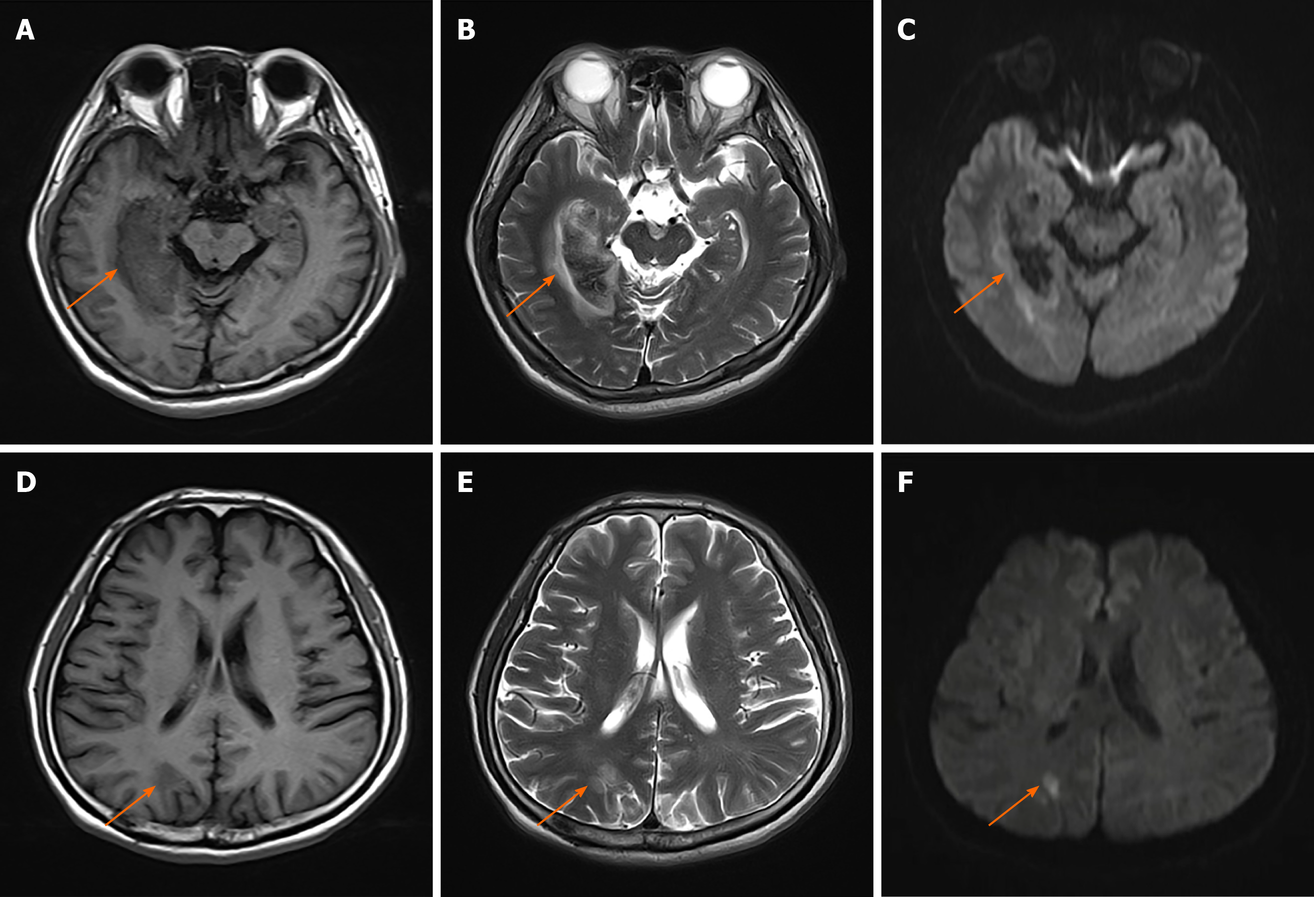
Figure 1 rain magnetic resonance imaging at admission.
A-C: Demonstrating a right occipital-temporal lobe hemorrhagic stroke. T1-weighted (A), T2-weighted (B), diffusion-weighted imaging (DWI) (C); D: T1-weighted presenting an occipital-parietal lobe ischemic stroke; E: T2-weighted presenting an occipital-parietal lobe ischemic stroke; F: DWI presenting an occipital-parietal lobe ischemic stroke. Orange arrowheads indicate the lesions.
- Citation: Yuan Y, Huang F, Gao ZH, Cai WC, Xiao JX, Yang YE, Zhu PL. Delayed diagnosis of prosopagnosia following a hemorrhagic stroke in an elderly man: A case report. World J Clin Cases 2020; 8(24): 6487-6498
- URL: https://www.wjgnet.com/2307-8960/full/v8/i24/6487.htm
- DOI: https://dx.doi.org/10.12998/wjcc.v8.i24.6487


