Copyright
©The Author(s) 2020.
World J Clin Cases. Dec 26, 2020; 8(24): 6450-6455
Published online Dec 26, 2020. doi: 10.12998/wjcc.v8.i24.6450
Published online Dec 26, 2020. doi: 10.12998/wjcc.v8.i24.6450
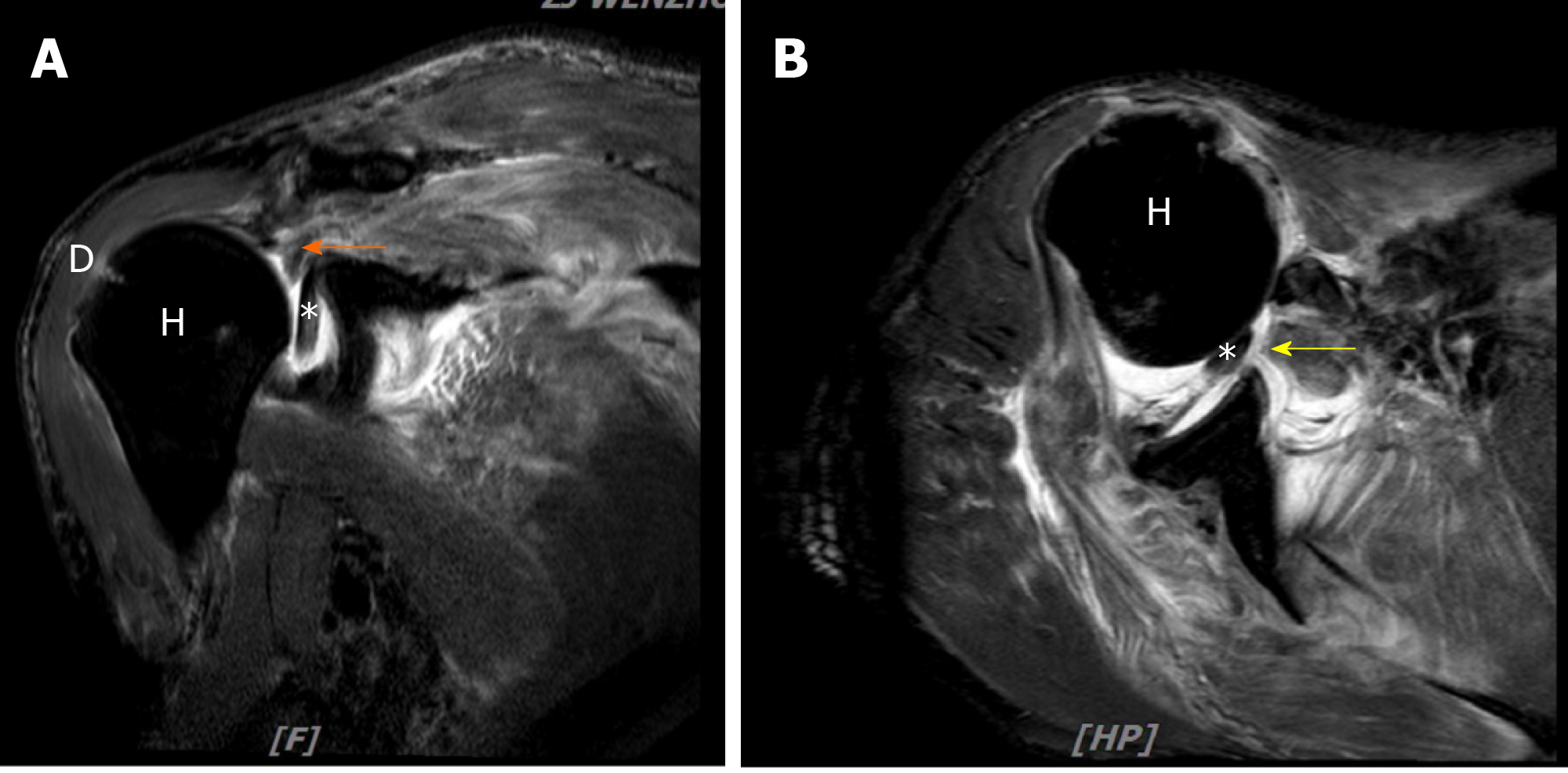
Figure 2 Magnetic resonance imaging of the right shoulder.
A: Coronal T2 fat-suppressed image; B: Axial T2 fat-suppressed image showing a full-thickness tear of supraspinatus, infraspinatus subscapularis tendons, and interposition of long head bicep tendon. The orange arrow indicates supraspinatus tendon, and the yellow arrow indicates subscapularis tendons. H: Humeral head; D: Deltoid muscle; Asterisk: Long head bicep tendon.
- Citation: Chen L, Liu CL, Wu P. Fracture of the scapular neck combined with rotator cuff tear: A case report. World J Clin Cases 2020; 8(24): 6450-6455
- URL: https://www.wjgnet.com/2307-8960/full/v8/i24/6450.htm
- DOI: https://dx.doi.org/10.12998/wjcc.v8.i24.6450


