Copyright
©The Author(s) 2020.
World J Clin Cases. Dec 26, 2020; 8(24): 6425-6431
Published online Dec 26, 2020. doi: 10.12998/wjcc.v8.i24.6425
Published online Dec 26, 2020. doi: 10.12998/wjcc.v8.i24.6425
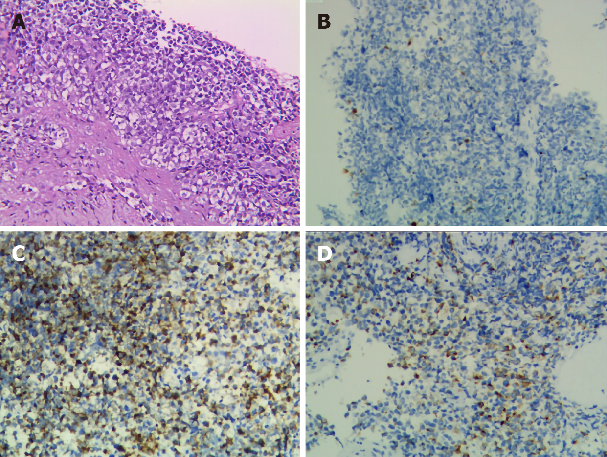
Figure 2 Pathologic results.
A: Representative images of tumor cells. Hematoxylin and eosin stain. Magnification: × 100; B: Immunohistochemical staining showed that the tumor was positive for S-100; C: For HMB-45; D: For melan-A.
- Citation: Long GJ, Ou WT, Lin L, Zhou CJ. Primary gastric melanoma in a young woman: A case report. World J Clin Cases 2020; 8(24): 6425-6431
- URL: https://www.wjgnet.com/2307-8960/full/v8/i24/6425.htm
- DOI: https://dx.doi.org/10.12998/wjcc.v8.i24.6425


