Copyright
©The Author(s) 2020.
World J Clin Cases. Dec 26, 2020; 8(24): 6373-6379
Published online Dec 26, 2020. doi: 10.12998/wjcc.v8.i24.6373
Published online Dec 26, 2020. doi: 10.12998/wjcc.v8.i24.6373
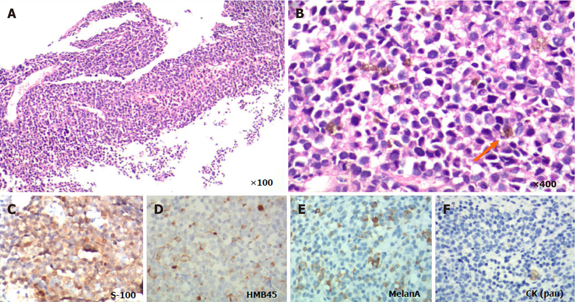
Figure 2 Microscopic examination of the tumor.
A and B: Hematoxylin-eosin staining showing the histological feature of the tumor cells. Fine melanin granules exited in or out of the cells (orange arrow); C-E: Immunohistochemical results shown positive staining of human melanoma black 45, S-100 and Melan A, and negative staining of cytokeratin (pan); F: In biopsy tissue (image C-F is magnified 400 ×).
- Citation: Xi JM, Wen H, Yan XB, Huang J. Primary pulmonary malignant melanoma diagnosed with percutaneous biopsy tissue: A case report. World J Clin Cases 2020; 8(24): 6373-6379
- URL: https://www.wjgnet.com/2307-8960/full/v8/i24/6373.htm
- DOI: https://dx.doi.org/10.12998/wjcc.v8.i24.6373


