Copyright
©The Author(s) 2020.
World J Clin Cases. Dec 26, 2020; 8(24): 6330-6336
Published online Dec 26, 2020. doi: 10.12998/wjcc.v8.i24.6330
Published online Dec 26, 2020. doi: 10.12998/wjcc.v8.i24.6330
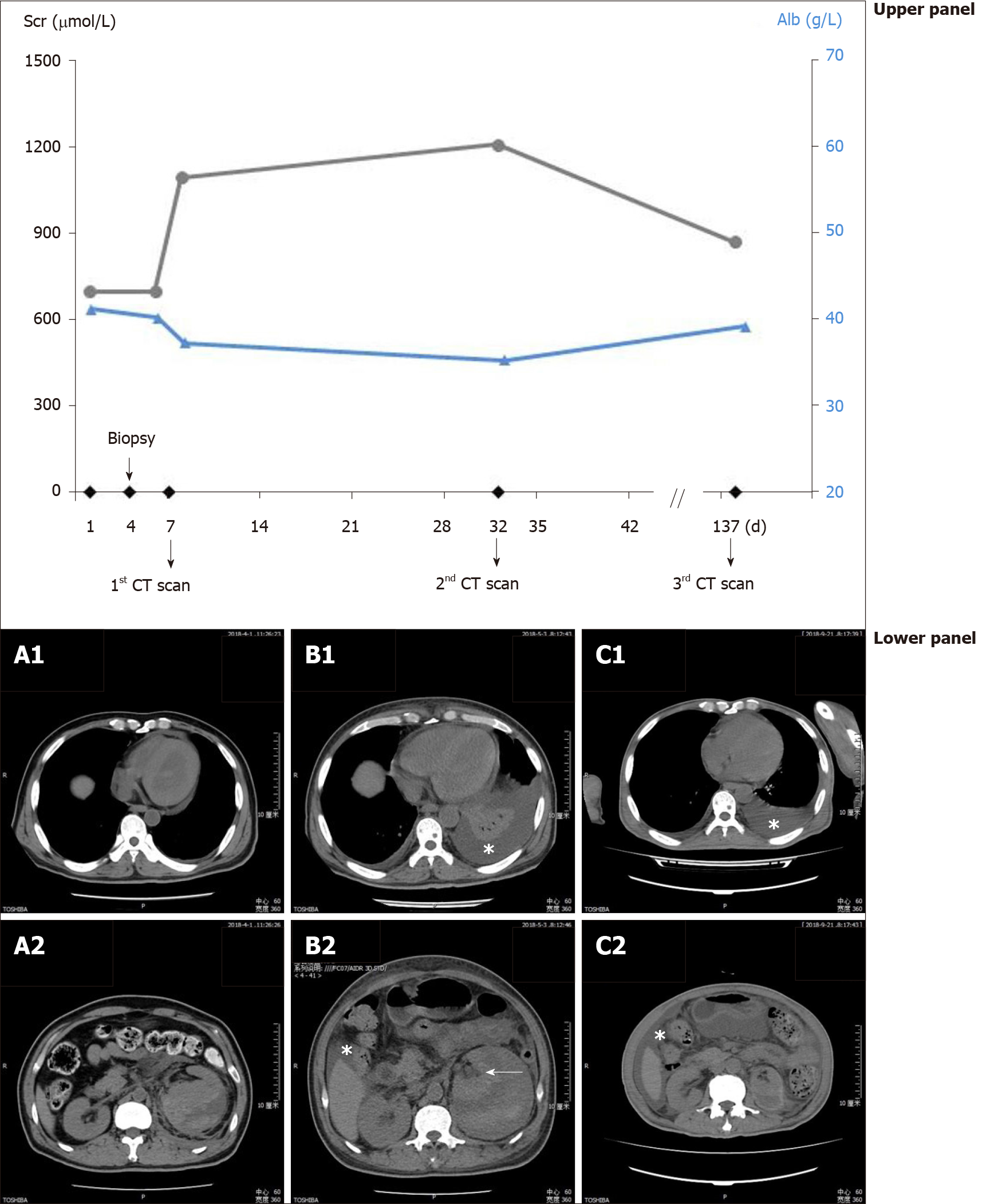
Figure 1 Evolution of the perirenal hematoma, pleural effusion and ascites in parallel with the corresponding serum creatinine and plasma albumin.
Upper panel: Values of serum creatinine (circles connected by black lines) and plasma albumin (triangles connected by blue lines) at different time points; Lower panel: A: Computed tomography scan shortly after the detection of hemorrhage showing perirenal hematoma (A2), without pleural effusion (A1); B: Perirenal hematoma and small amount of ascites (B2), with pleural effusion (B1, asterisk) 1 mo after the hemorrhage, the compressed kidney is also visible (B2, arrow); C: Perirenal fluid retention and ascites (C2), with pleural effusion (C1, asterisk) 5 mo after the hemorrhage. Alb: Albumin; CT: Computed tomography; Scr: Serum creatinine.
- Citation: Lin QZ, Wang HE, Wei D, Bao YF, Li H, Wang T. Pleural effusion and ascites in extrarenal lymphangiectasia caused by post-biopsy hematoma: A case report. World J Clin Cases 2020; 8(24): 6330-6336
- URL: https://www.wjgnet.com/2307-8960/full/v8/i24/6330.htm
- DOI: https://dx.doi.org/10.12998/wjcc.v8.i24.6330


